What to Expect at a Breast Clinic Appointment?
- Updated on: Jul 29, 2024
- 4 min Read
- Published on Nov 13, 2020

Breast Cancer
Breast cancer is a severe and most common problem observed in one in every eight women in the United States. Breast cancer leaves a great impact on any woman suffering from it and even on her caregivers. After breast cancer symptoms become prominent, women are referred to the breast clinic for proper breast cancer diagnosis. Detection and diagnosis should be the first priority without any delay.
What to Expect at Breast Clinic?
The symptoms do not lead to breast cancer always. In most cases, breast cancer is benign and non-cancerous, but it is always better to visit a breast cancer clinic and get a proper diagnosis through breast screening appointments. During the assessment, the doctors might ask about your health history. Several tests may be conducted for the diagnosis, making you feel nervous about the situation but do not feel anxious as a proper diagnosis is very important.
It is completely natural to feel worried and panic about the situation, but staying positive gives strength to combat the situation.
What Happens at a Breast Clinic?
In a breast clinic, any abnormality related to breasts is diagnosed. Multiple breast problems are analyzed, and accordingly, the treatment is provided to the patients.
Some specific tests or clinical examinations are conducted for breast cancer detection, such as:
- Mammogram
- Ultrasound scan
- Fine needle aspiration (FNA)
- Core biopsy
A combination of breast imaging (a mammogram and/or an ultrasound scan) and tissue sampling (a core biopsy or FNA) is known as a triple assessment that helps develop a definite diagnosis of cancer. A triple assessment test is used in severe cases when a definite diagnosis is imprecise.
Breast screening appointments are scheduled at the breast cancer clinic for women at risk of breast cancer. Breast screening is also done for women above 50 years once in every three years. It prevents the risk of breast cancer in aged-women and, if in some cases, there are chances of pre-breast cancer, proper treatment can be given to the patient at the right time.
What Happens at Breast Screening?
During the breast screening procedure, the doctor examines the breasts and the nearby areas (lymph organs-nodes) to detect any lump in the armpits or neck. A mammogram appointment is then given to the patient. Breast screening is done at a breast screening unit in a breast assessment clinic, and it may take about 20-30 minutes. Digital mammograms are very popular these days for the screening procedure.
Mammogram Test- What Happens at a Mammogram Appointment?
A mammogram is a breast X-ray and is considered the best way to detect early-stage breast cancer even before the symptoms might occur. Regular mammogram analysis lowers the risk of dying from breast cancer.
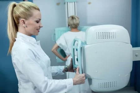
Mammogram test pictures soft tissue of the breasts in detail, which helps detect any abnormality. A mammogram technologist sets the position of the breasts one at a time between two special plates, and x-rays pass through them.
The breasts are flattened when the upper plate is compressed on the breasts for a high-quality picture. The upper plate, which is made up of plastic, is used to compress the breast (for a few seconds) while the technologist takes a picture. Mammogram analysis may take up to 30min, but breast compression may only last a few seconds.
Two or three images of each breast are taken from different angles for better analysis. Women below 35 are not screened using mammograms because breasts are denser at a younger age, which makes the X-ray images uncertain, and any changes are harder to identify. A mammogram report may be generated in 1 or 2 weeks after the screening.
Read About Breast Cancer and Genetics: BRCA Gene
Ultrasound- Another Technique for Breast Screening
High-frequency sound waves are used to produce images of breast tissues during an ultrasound. This is a no-pain and straightforward technique to detect breast cancer tumors, and there is no age-bar for this technique. During an ultrasound, a gel is spread over and around the breasts, which helps to get a clear picture. A blunt, thick pen or probe is made to move all around the breasts. A clear picture of the breasts’ inner tissues is seen during an ultrasound if there is no tumor present.
Fine Needle Aspiration (FNA) – Test to Confirm Image Analysis
For FNA, one or more samples of breast cells are collected using a fine needle and syringe. FNA test is conducted when any abnormality is detected in a mammogram test and in ultrasound pictures. The sample collected at the breast assessment clinic is sent for laboratory (microscopic) testing for confirmation. This test might be uncomfortable for many and might require the use of local anesthesia in some cases.
Core Biopsy- When FNA Is Not Efficient
Core biopsy is similar to FNA. A small part of tissue cells inside the breast is taken using a large needle. A small cut is made on the skin, and some tissue is extracted through it to obtain more detailed information. Local anesthesia is given to the patient at the breast cancer clinic before the procedure. Core biopsy is done when the lumps or tumors are needed to be studied more deeply.
In some cases, very specific pictures are required for detecting the exact location of the tumor cells. For this, the mammogram machine is linked to a computer. This is known as a stereotactic core biopsy.
After these tests, the doctor will study the results and tell the next step towards the treatment. If the result is not positive, then the doctor might suggest re-testing in some doubtful cases.
