Vitrectomy: Types, Procedure, Recovery
- Updated on: Jul 9, 2024
- 4 min Read
- Published on Oct 31, 2018


What is vitrectomy eye surgery?
Vitrectomy, which is also referred to as vitreous surgery is a type of eye surgery done to treat problems of the retina and vitreous of the eye. The procedure involves removal of vitreous humor gel or removal of blood or removal of scar tissue (that cause poor vision). This leads to providing a better access to the retina. It also allows a variety of repairs such as laser repair of retinal detachments, treatment of macular holes, etc.
Eye vitrectomy is recommended to the patients when the vitreous gel is infected, or an inflammation is observed, or the cavity is filled with blood or bits of tissues known as floaters. During the surgical procedure, vitreous gel (inside the eyeball) is replaced with saline water, a gas bubble or silicone oil. These fluids may be injected into the vitreous gel to help hold the position of your retina.
Generally, vitrectomies have a high success rate and vitrectomy cost depends on a surgeon’s expertise and the type of procedure. Vitrectomy with membrane peel is the most common vitreoretinal surgical procedure to remove epiretinal membrane (ERM). If ERM is not removed, it leads to the loss of foveal reflex, parafoveal light reflection, wrinkling of the retinal surface, etc.
Types of vitrectomy surgery
The surgery can be done in many different ways.
Posterior Pars Plana Vitrectomy (PPPV)
PPPV is a closed system procedure in which the vitreous humor is surgically removed through a needle inserted in the pars plana ciliaris. PPPV is performed for diseases of the posterior segment of the eye by a vitreoretinal surgeon.
Anterior Vitrectomy
During an eye trauma or injury, cornea or lens problem, in cataract or during cataract surgery after vitrectomy or during glaucoma surgery, vitreous gel comes out through the pupil in the anterior segment of the eye. In such cases, to avoid serious problems such as retinal detachment and glaucoma, vitreous is removed with the anterior approach for early recovery.
Need for vitrectomy operation
- Diabetic retinopathy leads to the bleeding or scarring of tissues which affects the retina. This means that the retina is damaged by long period of uncontrolled diabetes.
- Vitreous haemorrhage: If bleeding occurs in the vitreous gel, it can lead to clouding or blurry vision. Serious eye injuries may result in a vitreous haemorrhage.
- Retinal detachment: If the retina separates from the back of the eye cavity, the vitreous gel or fluid leaks through a retinal tear or hole. This collects under the retina and causes it to separate from the tissue around it. Retinal detachment surgery is recommended for this problem.
- Macular degeneration: Macular hole or a tear in the centre of the retina (or macula) is observed in the eye which causes loss of reading and detailed vision. This condition is called macular degeneration.
- Vitreoretinal traction: It generally occurs due to aging, nearsightedness, severe diabetes, severely premature infants, head or eye trauma which leads to abnormal pulling on the retina.
- Excessive scaring of tissue occurs on the surface of the retina and in the vitreous. It can pull on the retina, lifting it up like a tent and puckering it into stiff folds. Scarring leads to distortion, blurring or double vision.
- In some rare cases, during a cataract surgery, the natural lens of the eye falls into the vitreous cavity which results in inflammation and an increase in the eye pressure. With the help of vitrectomy, the dislocated lens can be removed.
- Infection in the eye, called endophthalmitis, occurs due to an eye surgery or due to any foreign object stuck inside the eye from any injury. This can also be treated through vitrectomy procedure.
- Macular pucker (wrinkles or creases in the macula) also leads to distorted vision problems.
- Vitrectomy for floaters is a very safe option for treating flash light problems.
Vitrectomy procedure
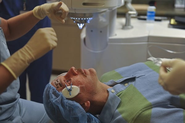
Vitrectomy operation is performed under either a local or general anesthetic. The eyelids are completely opened and three tiny openings are made in the sclera. Delicate instruments such as vitreous cutters, forceps and scissors help in removing the vitreous gel and scar tissues that grow on the surface of the retina. A fiber-optic light is inserted into one of the cuts to see through the inside of the eye.
The vitreous is removed and is replaced with another substance, such as gas bubble (vitrectomy gas bubble procedure), air, or a saline solution. These fluids will be replaced by a fluid that eye naturally produces over time. In case silicone oil or heavy oil is used as a vitreous substitute to heal serious conditions, another surgical procedure is needed several months after the surgery for the removal of silicone oil.
Laser vitrectomy is another popular surgery to repair the retina or to remove damaged tissue from the eye. A fibreoptic illuminator is used to light the inside of the eye during the operation.
Vitrectomy recovery
In general, vitrectomy recovery time is estimated to be between 4 to 6 weeks. There are chances of temporary mild swelling on the eyelids, bruising around the eye, and redness following the surgery which improves relatively quickly. Severe pain or sensation is very uncommon unless there is unusual inflammation or high eye pressure.
Eye drops are prescribed by doctors to help avoid infection and reduce the inflammation. Several pain medications, such as nonsteroidal anti-inflammatory drugs (NSAIDs) like ibuprofen (Advil) help in pain management or soreness in the eyes. In a few cases, doctors recommend wearing an eye patch for a few days to avoid any type of infection and increased pressure.
Risks & complications of vitrectomy surgery
Although, complications are rarely observed after vitrectomy surgery but it depends on the condition of the eye. There are few but possible chances of risks such as:
- Cataracts
- Retinal detachment
- Raised pressure inside the eye
- Infection or bleeding or fluid discharge in some cases
- Problems during eye movement
- Damage to eye lens (rarely)
- Loss of vision after vitrectomy (rarely)
- Floaters or flashes of light


