Valvular Heart Disease: Diagnosis and Management
- Updated on: Nov 1, 2023
- 4 min Read
- Published on Aug 26, 2022
Any heart valve that has been damaged or is ill is said to have valvular heart disease. Valve illness has a number of causes. Right and left atria, as well as right and left ventricles, make up the four chambers and four valves of a typical heart.
- The bicuspid or mitral valve, which is located between the left atrium and left ventricle, permits blood to pass from one chamber to the other.
- Blood can move from the right atrium to the right ventricle thanks to the tricuspid valve.
- Blood can move from the left ventricle to the aorta thanks to the aortic valve.
- Blood can move from the right ventricle to the pulmonary artery through the pulmonary valve.
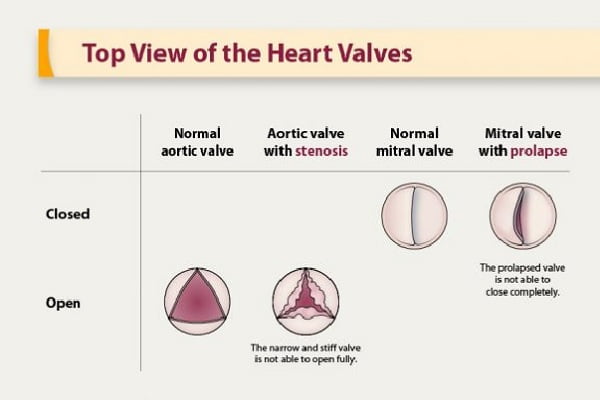
The blood flow into and out of the heart is regulated or controlled by the valves opening and closing. The three leaflets or flaps that make up three of the heart valves work together to open and close the valves, allowing blood to flow through the opening. There are just two leaflets in the mitral valve. When the heart beats, leaflets on a healthy heart valve can fully open and close the valve, but diseased valves may not. The aortic valve is the most often afflicted heart valve, yet any valve might develop a problem. Regurgitation is a condition where a diseased valve partially closes, or becomes “leaky.”
If this takes place, blood seeps back into the original chamber and the heart is unable to pump enough blood forward. The other typical heart valve disorder is stenosis, which occurs when the valve’s aperture is constricted and stiff and the valve is unable to fully open while blood is trying to pass through.
Sometimes a valve’s leaflet may be absent; the aortic valve is more frequently affected by this. If the heart valves are damaged, the heart must work harder to pump blood throughout the body, either against a restricted aperture or as blood leaks back into the chamber.
Diagnosis
During the physical examination, your doctor will listen for a heart murmur, which could be an indication of a problem with the heart valves. Several tests could be used to diagnose your disease.
Tests could consist of:
Echocardiography:
Video images of your beating heart are created when sound waves from a wand-like instrument (transducer) are aimed at your heart. With this exam, your heart’s anatomy, heart valves, and blood flow are evaluated. Your doctor can examine the heart valves closely and determine how well they are functioning with the aid of an echocardiography. Additionally, doctors might employ a 3D echocardiography.
A transesophageal echocardiography is an additional type of echocardiogram that involves inserting a tiny transducer at the end of a tube down the tube going from your mouth to your stomach (esophagus). With this test, your doctor can examine the heart valves more closely than they could with a standard echocardiogram.
Electrocardiogram (ECG):
Electrical impulses from your heart are measured by wires (electrodes) placed to pads on your skin. An ECG can identify cardiac illness, enlarged heart chambers, and irregular heart rhythms.
A chest X-ray:
Your doctor can evaluate whether the heart is enlarged, which may suggest specific types of cardiac valve problems, with the aid of a chest X-ray. The condition of your lungs can also be determined by doctors using a chest X-ray.
Heart MRI:
Using magnetic fields and radio waves, a cardiac MRI can produce precise images of your heart. It can examine your condition’s severity and the size and efficiency of your lower heart chambers.
Stress testing or exercise tests:
Various exercise tests can be used to gauge your activity level and track how your heart reacts to physical stress. If you are unable to exercise, you may be prescribed drugs that imitate the positive effects of exercise on the heart.
Catheterization of the heart:
This test isn’t frequently used to diagnose heart valve disease, but it might be if other tests can’t do so or can’t tell how severe the condition is.
In order to see an artery in your heart on an X-ray, a doctor would thread a thin tube (catheter) into a blood vessel in your arm or groin, direct it to the artery, and inject dye through the catheter. This gives your doctor a thorough image of your heart’s arteries and how it works. Additionally, it has the ability to gauge heart chamber pressure.
Treatment
Treatment for heart valve disease is based on your symptoms, the severity of your condition, and if it is becoming worse. Your care will be given by a physician (cardiologist) with specialized training in heart disease. Monitoring your condition through many follow-up visits may be part of the treatment. You could be requested to:
- Change your lifestyle for the better.
- To treat symptoms, take medications.
- If you experience atrial fibrillation, a specific type of abnormal heart rhythm, take blood thinners to lower your risk of blood clots.
Procedures such as surgery
Even if you don’t have any symptoms, you may eventually require heart valve surgery to replace or repair the damaged heart valve. Your doctor may decide to replace or repair the damaged valve at the same time as any surgery required to treat another heart ailment. The typical method of performing heart valve surgery is through a cut (incision) in the chest. Minimally invasive heart surgery, which requires fewer incisions than open heart surgery, is occasionally performed by doctors. Robot-assisted heart surgery is a sort of minimally invasive cardiac surgery where physicians employ robotic equipment to carry out the process in specific medical facilities.
There are surgical options for replacing or repairing valves:
Heart Valve Replacement
If the injured valve cannot be healed, surgeons may replace it with a mechanical valve or a valve manufactured from cow, pig, or human heart tissue after removing the damaged valve (biological or tissue valve). If a mechanical valve was used to replace your natural valve, you will always need to take blood thinners to avoid blood clots. Over time, biological tissue valves deteriorate and typically need to be replaced.
A damaged aortic valve may be replaced using a minimally invasive method known as transcatheter aortic valve replacement (TAVR). A long, thin tube (catheter) is inserted into an artery in your leg or chest during this treatment, and it is then directed to the heart valve.
Heart Valve Repair
To keep your heart valve healthy, your doctor might advise heart valve repair. During a heart valve repair, doctors may:
- Repair valve leaking areas
- Fused leaflets of a separate valve
- Replacing the valve support cables
- To enable the valve to tightly seal, trim any extra valve tissue.
The ring around a valve (annulus) is frequently tightened or strengthened by surgery by implanting an artificial ring. In rare instances, doctors employ long, thin tubes and less intrusive techniques to repair specific valves (catheters). Clips, plugs, or other tools may be used during these procedures.
