A Quick Eye Scan Can Now Detect Alzheimer’s
- Updated on: Jul 2, 2024
- 2 min Read
- Published on Mar 26, 2019
Diagnosing Alzheimer’s disease is a big challenge as there is no specific and known method to identify this particular problem. There are various possibilities being explored to fight and overcome this challenge.
In the current scenario, a known invasive method is supposedly the only way to diagnose Alzheimer’s disease. This non-invasive method is a kind of lumbar puncture or spinal tap method during which some cerebrospinal fluid is drawn out of the body with the help of a needle inserted in the back and a PET scan is then conducted to take images of the amounts of beta-amyloid, a toxic protein which accumulates in Alzheimer’s disease.
Alzheimer’s and its extent
Alzheimer’s is a neuro-cognitive disease which results in loss of memory, loss in the ability to judge, to function, to learn, etc. It is supposed to be a growing catastrophic event for public health. A study in 2014 estimated that more than 5million people in the United States suffer from Alzheimer’ disease and is the sixth leading cause of death in America.
A study stated that in Louisiana only about 87,000 people are affected with Alzheimer’s disease, which is likely to increase to about 110,000 till 2025. According to the CDC, the death rate due to Alzheimer’s has elevated by more than 50 percent, recently. Georgia Health News stated that the casualty number due to Alzheimer’s has increased by 201 percent since 2000.
In the United States, November is referred to as Alzheimer’s Awareness Month since 1983. This designation was given by President Ronald Reagan.
Spot Alzheimer’s disease through an eye scan: Is it possible?
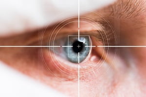
There are some suggested studies which indicate that Alzheimer’s disease can be diagnosed with the help of an eye scan. Earlier, brain imaging tests such as MRI scans, CT scans, etc or neuro-physiological testing were the only options available to identify Alzheimer’s disease but these methods cannot be completely trusted for accurate results.
Certain trial and tested methods have indicated a hopeful ray of light that a new kind of non-invasive imaging technique known as optical coherence tomography angiography (OCTA) focuses on tracking the connection of between the eye and the Alzheimer’s disease. This study stated that the retina (present at the back of the eye) of Alzheimer’s patients is diagnosed with mild cognitive impairment.
The theory for this technique states that the retina, which joins the brain with the help of an optic nerve, faces deterioration. The retinal blood vessels help in viewing the changes going on in some structures and blood vessels in the brain. This helps to locate any changes in the brain and its structures that are evident to prove Alzheimer’s syndrome.
In another study, the images of brain and retina demonstrated that the retina gets thinner with time and the hippocampus becomes smaller which ultimately leads to loss of mental abilities. This is a sign of Alzheimer’s disease and is observed especially in those people who have had some family history of either Alzheimer’s or dementia.
Why is there a need of an evolved diagnostic method to detect Alzheimer’s?
Lumber puncture method is an invasive way of detecting Alzheimer’s and may or may not give proper results. This method is expensive and the disease is detectable at a very later stage. Lumber puncture method is insufficient for screening many patients.
Therefore, various researches suggested that early neurodegeneration can be predicted in the eye through an eye scan. This is related to the thinning of the retina and also indicates the onset of Parkinson’s disease. A quick, cheap and painless eye scan can be a revolutionary innovation if proved to be about 100% accurate.
