Stomach Cancer – Definition and Overview
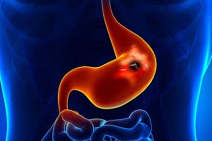
The growth of a stomach cancer (also called gastric cancer) begins when cancerous cells start developing in the grandular tissues of the inner mucus lining of stomach. Stomach cancers have a tendency to grow slowly over the years. Early changes in the mucosal lining of the cells rarely shows any symptoms and therefore often go undetected leading to delayed diagnosis and treatment of the stomach cancer.
Stomach cancer is also known as gastric cancer in many countries, such as in Japan, China, Chile, and Iceland.
According to the World Health Organization (WHO), out of all cancer-related deaths worldwide 723,000 are caused by stomach cancer every year. It is the fifth most common cancer worldwide, but the third leading cause of cancer-related deaths.
The National Cancer Institute (NCI) estimated that approximately 28,000 new cases of stomach cancer were found in 2017.
The most commonly occurring type is “adenocarcinoma” of the stomach. In an adenocarcinoma, cancerous cells develop in the mucosal lining, which is the most superficial but inner lining of the stomach and helps produce mucus.
Other less common types of gastric or stomach cancers are gastrointestinal carcinoid tumors (small cell carcinomas), neuroendocrine tumors, gastrointestinal stromal tumors, and lymphomas.
If the stomach cancer is not detected and treated at an early stage, it can extend to the lymph nodes of the lymphatic system or to other organs in the body through bloodstream, such as to the liver and lungs.
Stomach cancer slowly expands to the walls of the abdomen (peritoneum) and sometimes also grows through the stomach wall into nearby organs such as the pancreas and bowel.
About 50% of the stomach cancers occur in people with age of about 65 or over.
More: Graphics, images, and photographs for stomach cancer (gastric cancer)
The stomach
The stomach is a hollow muscular sac-like organ which is an essential part of the digestive system in our body. The stomach holds the food and the digestion begins here with the secretion of gastric juices (produced by the glands in the inner lining of the stomach). After this, the semi-solid mixture of the food and gastric juices is poured into the deudenum (first part of the small intestine).
Mechanism of food digestion
The mechanism of food digestion begins when the food enters the esophagus (a tube that carries food through the throat and chest to the stomach). The esophagus extends below in downward direction and meets the stomach at the gastroesophageal (GE) junction, just beneath the diaphragm (the thin sheet of breathing muscle under the lungs). The food is then broken down into a semi-solid mixture that then passes into the small intestine.
The stomach is placed in the upper abdomen of the body and mainly has three main parts. They are:
- The upper part, which is divided into cardia and fundus which helps to prevent stomach contents from going back up into the esophagus.
- Middle part is the “body” (corpus) of the stomach between the upper and lower parts where food is mixed and its breaking down starts.
- Bottom portion comprises of the antrum and pylorus which acts as a valve that controls the emptying of the stomach contents into the small intestine. It holds the broken-down food until it is ready to be released into the small intestine. It is sometimes called the pyloric antrum.
The upper part of the stomach which mainly comprises of cardia, fundus, and the body are called proximal stomach. Sometimes the cells in the upper part of the stomach make acid and pepsin (a digestive enzyme) and also the gastric juices that help in food digestion.
The lower 2 parts which comprise the antrum and pylorus are known as the distal stomach. The two curves of the stomach forms its inner and outer borders, and are called the lesser curvature and greater curvature, respectively.
The stomach wall has 5 layers:
- The innermost layer of the stomach is the mucosa (mucosal membrane) where stomach acid and digestive enzymes are produced. Most stomach cancers begin from mucosa (a ridge shape structure).
- It is then aligned with a supporting layer called the submucosa. The submucosa contains larger blood and lymph vessels, nerve cells and fibres.
- These two layers are covered by the muscularis propria. The movement in this layer helps in mixing stomach contents.
- The outer 2 layers, composed of the subserosa and the outermost serosa, wrap the stomach. The serosa is the fibrous membrane that covers the outside of the stomach. The serosa of the stomach is also called the visceral peritoneum.
Growth and progression of stomach cancer
The tumor cells in the stomach grow slowly and behave normally without showing any signs and symptoms of the cancer in the beginning. These tumors can be sometimes non-cancerous or benign tumours such as gastric polyps, small gastrointestinal stromal tumours (GISTs) or lipomas etc.
Stomach cancer develops slowly over a period of several years after the cells start growing at a high and uncontrolled rate. Cancerous cells may grow at different sections of the stomach causing manifestation of different types of symptoms at different levels. In different stages, the cancer tends to have different prognosis.
The location of your stomach cancer also affects your treatment. For example, cancers that begin at the gastroesophageal junction (GE junction) are usually treated the way cancers of the esophagus are treated and managed. A cancerous tumor that mainly begins in the cardia of the stomach is also staged and treated like a cancer of the esophagus because the cells multiply into the GE junction.
During a pre-cancerous stage, changes often occur in the inner lining (gastric mucosa) of the stomach but the cells are not malignant at this stage and early changes rarely manifest into symptoms. Therefore, stomach cancer often goes undetected in the early stages. Pre-cancerous conditions that can develop into stomach may include such as gastric adenoma, or adenomatous polyps, and gastric epithelial dysplasia.
More: Colorectal Cancer – Overview and Types
More: Anti–Vascular Endothelial Growth Factor Therapy (Anti VEGF Therapy) for Cancers
Types of stomach cancers
Adenocarcinoma
Most of the stomach cancers (more than 85%) are adenocarcinomas. Adenocarcinoma develops from the cells that form the innermost-mucosal lining of the stomach.
Adenocarcinoma is classified into two main sub-categories:
Intestinal adenocarcinoma
The cells in the intestinal adenocarcinoma are arranged as tubes similar to the mucosa of the intestines. The cells of intestinal adenocarcinoma are well differentiated most of the times, which means the cells appear and behave like normal cells.
Intestinal adenocarcinoma is of three types:
- tubular adenocarcinoma
- papillary adenocarcinoma
- mucinous adenocarcinoma.
Intestinal adenocarcinoma is more common in men than in women.
Diffuse adenocarcinoma
Diffuse adenocarcinoma is inclusive of infiltrative, mucinous and signet ring carcinoma. Diffuse adenocarcinoma is undifferentiated cancer cells which are scattered throughout the stomach lining and do not appear as normal cells. Diffuse adenocarcinoma is more common in people with comparatively lower age.
In the infiltrative type of diffuse adenocarcinoma, there is no formation of any mass or lump or ulcer. It often leads to “linitis plastic” where the stomach wall becomes hard and leathery.
Other, rare types of adenocarcinoma
- mixed adenocarcinoma (both intestinal and diffuse adenocarcinoma)
- gastric lymphoepithelioma-like carcinoma (LELC)
- hepatoid adenocarcinoma
Lymphoma
Gastric lymphomas are generally non-Hodgkin lymphomas (NHL). Gastric cancers (of the immune system tissue) can sometimes be found in the wall of the stomach. The treatment and outlook depend on the type of lymphoma.
Mucosa-associated lymphatic tissue (MALT) lymphoma is the most commonly occurring type of gastric lymphoma. Mostly, gastric MALT lymphoma occurs due to H. pylori (Helicobacter pylori) infection which begins with the formation of ulcer. The treatment for gastric MALT lymphoma associated with H. pylori involves the use of antibiotics and proton-pump inhibitors.
Gastrointestinal stromal tumor (GIST)
GISTs are very rare and aggressive tumors in early forms of cells in the wall of the stomach known as the interstitial cells of cajal. These tumors are very slow growing. Some of these tumors are non-cancerous (benign) while the others which are cancerous are known as carcinoid tumors.
Although GISTs can be found anywhere in the digestive tract (most are found in the stomach). They are sometimes called carcinoid tumors. Treatment of GISTs involves surgery or biological therapy. Many types of biological therapies may be employed for this purpose such as somatostatin cogener therapy. In a somatostatin cogener therapy, certain chemicals that mimic somatostatin, a hormone that prevents the release of other hormones, are used.
Carcinoid tumor
A carcinoid tumor begins in hormone-producing cells of the stomach. Most of these tumors do not expand to other organs.
Adenosquamous carcinoma involves a mix of features of both adenocarcinoma and squamous cell carcinoma. The outlook of adenosquamous cell carcinoma tends to be less favorable than adenocarcinoma.
Other rare stomach cancers
Other rare types of cancers that can begin in the stomach are:
- squamous cell carcinoma
- neuroendocrine tumors
- small cell carcinoma
- leiomyosarcoma
Key statistics about stomach cancer
Stomach cancer mostly affects old age people. The average age of people when diagnosed with stomach cancer is found to be about 68. According to the American Cancer Society, about 60% of people who were diagnosed with stomach cancer in a year were 65 years in age or older. The chances of getting stomach cancer in a person’s lifetime are about 0.01% in men and 0.065 in women.
The prognosis of stomach cancer is estimated about 42%, which means about 42 in 100 people survive for at least one year after the diagnosis. 19% of the people diagnosed with stomach cancer live for at least five years after the diagnosis. Only 15% survive for at least 10 years after the diagnosis.
More: Salivary Gland Cancer: Causes, Signs, Symptoms, Diagnosis, Treatment, Prevention, Stages
Stomach Cancer Risk Factors
The risk of stomach cancer increases with an increase in age. It most commonly affects men, though women can also get it. Some known risk factors of stomach cancer, which seem to play a significant role in the occurrence of stomach cancer, are:
Helicobacter pylori infection
The most important risk factor for stomach infection is supposed to be Helicobacter pylori infection. Helicobacter pylori infection is considered the major cause of stomach cancer, especially if the infection occurs in the lower part of the stomach.
H. pylori is a common type of bacteria found in the stomach which can cause inflammation (called chronic atrophic gastritis). Ulcers and pre-cancerous changes to the cells in the lining of the stomach begin after the inflammation. Infection with Helicobacter pylori generally occurs in every 1 of 3 people suffering with stomach cancers.
While H. pylori can increase the risk, it is not necessary that all people who get infected with H. pylori will suffer from stomach cancer. H. pylori may act with other factors too and increase the overall risk of developing the stomach cancer. The age at which one is infected with stomach cancer may also increase the risk of someone developing the stomach cancer.
Age and sex
Stomach cancer is more common in older people. Approximately, 60% of stomach cancers develop in people aged 75 or over.
Men are nearly twice as likely to get the disease compared to women.
Tobacco intake
Smoking or intake of tobacco through any other means increases the risk of stomach cancer (particularly cancers in upper portion of the stomach near the esophagus). About 1 in 5 cases of stomach cancers is due to smoking or intake of tobacco. Tobacco doubles the stomach cancer chances.
Diet
A high risk of stomach cancer is observed in people whose diet regularly includes smoked food, salted fish and meat, and vegetable pickles.
Processed meat such as ham, bacon, salami and sausages if eaten in large quantities can increase the risk of stomach cancer. Nitrates and nitrites are substances commonly found in cured meats. These substances are converted by certain bacteria, such as H pylori, into cancer-causing compounds in the stomach.
Consuming fresh fruits and vegetables appear to lower the risk of stomach cancer.
Consumption of salty foods can increase the risk of stomach cancer. It risks around 1 in 4 stomach cancer cases.
Pernicious anemia
Certain cells in the stomach lining normally produce a substance called intrinsic factor (IF) for the absorptions of vitamin B12 from foods. The deficiency of Vitamin B12 strongly affects our body’s ability to manufacture new red blood cells. This condition is known as pernicious anemia. People with pernicious anemia have an increased risk of getting stomach cancer.
Menetrier disease (hypertrophic gastropathy)
Excessive growth of the stomach lining can result in large folds in the lining which can lead to lower level of stomach acid. Large folds occur because the cells in the lining of the stomach grow too much. The exact mechanism is unknown but this disease increases the chances of someone getting stomach cancer.
Inherited cancer syndromes
- Hereditary diffuse gastric cancer (HDGC): HDGC is caused by a mutation (defect) of the CDH1 gene (also known as E-cadherin gene). The cancerous cells start spreading throughout the stomach without forming a tumor at a single place. The stomach cancer risk among the affected people is about 70% to 80%.
- Familial adenomatous polyposis (FAP): Familial adenomatous polyposis (FAP) is mainly caused by a mutation of the adenomatous polyposis coli (APC) gene. Most polyps develop on the lining of the colon and rectum. Polyps can also develop in the stomach and small intestine. It also greatly increases the risk of colorectal cancer.
- Lynch syndrome: Lynch syndrome (also known as hereditary non-polyposis colorectal cancer or HNPCC) is caused by mutations in the genes that correct the problems when cells divide and the DNA is copied. The genes that can cause Lynch syndrome include MLH3, MSH6, TGFBR2, PMS1, and PMS2.
- Peutz-Jeghers syndrome: Peutz-Jeghers syndrome is caused by a mutation of the STK11 gene (also called PJS or LKB1 gene). It causes polyps (hamartomas) in the intestines and stomach.
- Li-Fraumeni syndrome: Li-Fraumeni syndrome is usually caused by mutations in the TP53 or CHEK2 gene. This syndrome is associated with a higher risk of developing several types of cancer, including stomach cancer, at a relatively young age.
- BRCA gene mutations: These mutations increase the risk of developing breast cancer or ovarian cancer. People who carry mutations of the inherited breast cancer genes BRCA1 or BRCA2 may also be associated with an increased risk of stomach cancer.
Other risk factors of stomach cancer
- A previous surgery which results in reflux (backup) of bile from the small intestine into the stomach after surgery adds to the risk of stomach cancer.
- Being overweight or obese can also put you at an increased risk of cancers of the cardia (the upper part of the stomach nearest the esophagus).
- People with type-A blood have a higher risk of stomach cancer.
- Exposure to fumes and dust particles at workplaces can increase the risk of developing stomach cancer, especially for people working in the rubber production industry.
- The risk of stomach cancer is 2 to 10 times higher in case of family history of the stomach cancer.
- Stomach cancer is very common in certain countries. It is more prevalent in China, some parts of Europe and America, and Japan. It is less common in Northern and Western Africa, South Central Asia, and North America.
Causes of stomach cancer
We don’t know what causes most stomach cancer cases. But there are some factors that may increase the risk of developing it. These are called stomach cancer risk factors and have been discussed above in the article.
Scientists believe that there are certain things related to the risk factors that may trigger or cause the stomach cancer. These are discussed here.
The normal glands of your stomach may be decreased. Some inflammation may also occur as the stomach cells are damaged by cells of your immune system if you have chronic atrophic gastritis. A person’s immune system generates autoimmune response attacking the cells lining the stomach. Atrophic gastritis occurs due to H. pylori infection. This might cause cancerous growth in the stomach.
Another possible cause of stomach cancer is pre-cancerous change in intestinal metaplasia. The normal lining of the stomach is replaced with cancerous cells that closely resemble the cells which usually line the intestine. This might also be related to H pylori infection.
Gastroesophageal junction cancer is often linked with having gastrointestinal reflux disease (GERD), and less commonly with obesity and smoking. GERD is a condition caused by frequent backflow of stomach acid into the esophagus which often occurs after a surgery.
Signs and Symptoms of Stomach Cancer
Early-stage stomach cancer rarely causes symptoms. This is one of the reasons, stomach cancer is often hard to detect early. But, if at all the symptoms appear, these may include:
- Reduced appetite
- Continuous weight loss
- Abdominal (belly) pain
- Stomach pain
- Buildup of fluid in the abdomen (called ascites) and swelling
- A lump in the abdomen that can be felt during a physical exam
- Wart-like growths on the skin (seborrheic keratoses)
- Vague discomfort in the abdomen, usually above the navel
- A lump in the area of the belly button, or navel (a swollen lymph node, sometimes called Sister Mary Joseph node)
- A sense of fullness in the upper abdomen after eating a small meal i.e. feeling bloated
- Severe, persistent heartburn or indigestion
- Unexplained, persistent nausea
- Vomiting, with or without blood
- Blood in the stool
- Low red blood cell count (anemia)
- Jaundice (the skin and whites of the eyes become yellow and urine is dark yellow)
- A lump on the ovary (Krukenberg tumour)
- A lump in the pelvis (Blumer shelf), which may be felt during a rectal exam
- A lump in the left armpit (a swollen lymph node)
- A lump above the left collar bone (1 or more swollen lymph nodes, sometimes called Virchow node)
- Darkening of the skin on body folds and creases (acanthosis nigricans)
Can Stomach Cancer Be Prevented?
To reduce the risk of gastro-esophageal junction cancer and stomach cancer, certain preventive measures can be taken, as follows. However, it is not possible to prevent the cancer completely because the exact cause of the stomach cancer is unknown.
Diet, nutrition, body weight, and physical activity for preventing stomach cancer
- A healthy diet reduces the risk of stomach cancer
- High in smoked and pickled foods and salted meats and fish should be avoided
- A diet high in fresh fruits and vegetables can also lower down stomach cancer risk. Citrus fruits (such as oranges, lemons, and grapefruit) may be especially helpful.
- Combinations of antioxidant supplements (vitamins A, C, and E and the mineral selenium) might reduce the risk of stomach cancer in people with poor nutrition.
- Choosing whole-grain breads, pastas, and cereals instead of refined grains, and eating fish, poultry, or beans instead of processed meat and red meat may help to lower the risk of cancer.
- Being overweight or obese may add to the risk of stomach cancer. Bbeing physically active may help lower your risk.
Avoiding tobacco use
- The consumption of tobacco increases the risk of cancers of the proximal stomach (the portion of the stomach closest to the esophagus).
- Tobacco use while smoking increases the risk for many other types of cancers; and therefore its use should be avoided.
The use of Aspirin
- The use of aspirin or other non-steroidal anti-inflammatory drugs (NSAIDs) such as ibuprofen or naproxen, sometimes prevent stomach cancer.
- These drugs can also reduce the risk of developing colon polyps and colon cancer.
- The disadvantage of using such medicines is that they can also cause serious (and even life-threatening) internal bleeding and other potential health risks in some people.
Treating H pylori infection
- Some specific antibiotics reduce the H pylori infection, which in turn lowers the number of pre-cancerous lesions in the stomach and reduce the risk of developing stomach cancer.
- The H pylori infection is detected through different tests such as breast test, biopsy or endoscopy and treated to reduce the chances of cancer development in the stomach.
Early detection and screening: Can Stomach Cancer Be Found Early?
Stomach cancer is an uncommon type of cancer. But often it is distinguished geographically, for example, stomach cancer is very common in Japan.
Mass screening of the population helps in finding many cases at an early and easily curable stage. Screening means testing people for early stages of an illness before they experience any symptoms.
In the US, routine screening for stomach cancer is generally not done, which results in most people not getting stomach cancer diagnosed until they have certain signs and symptoms that points to the need for medical tests. But, by this time the cancer has already spread to other locations and is difficult to cure, in most cases.
Diagnostic Tests for Stomach Cancer
Medical history and physical exam
Health history is a record of symptoms, risk factors and all the medical events and problems that a person has been associated with. During a physical exam, the signs of stomach cancer are observed.
The common signs that your doctor will look for during a physical examination are:
- enlarged organs, lumps or fluid in the abdomen
- swollen lymph nodes in the armpits and above the collar bone
- a digital rectal exam to check for a lump in the pelvis area
Upper endoscopy and endoscopy ultrasound
Upper endoscopy (also called esophagogastroduodenoscopy or EGD) is the common test used to detect stomach cancer. It is also known as Upper gastrointestinal (GI) endoscopy.
A thin, flexible, lighted tube with a small video camera on its end is inserted down the throat. The lining of the esophagus, stomach, and first part of the small intestine (duodenum) is examined. It may also be called a gastroscopy when only the stomach is examined.
Upper endoscopy helps in examining the stomach for bleeding, ulcers, polyps, tumors and inflammation (gastritis).
An ultrasound may be done with an endoscope (endoscopic ultrasound, or EUS) to detect how far the tumor has grown into the wall of the stomach and nearby tissues. EUS can also show the cancer that has spread to the nearby lymph nodes. In an endoscopic ultrasound (EUS), a small transducer is placed on the tip of an endoscope. The picture quality is better in EUS than in a standard ultrasound (because of the shorter distance for the sound waves to travel).
Endoscopy is used as part of a special imaging test known as endoscopic ultrasound. Through an endoscope, stomach cancer appears like an ulcer in shape of a mushroom or like a protruding mass, or like a flat, diffused structure with thickened areas of mucosa known as linitis plastic. It can also be used to guide a needle into a suspicious area to get a tissue sample (EUS-guided needle biopsy) and check for cancer in the wall of the stomach, outside the stomach or in surrounding lymph nodes.
Biopsy for stomach cancer diagnosis
The tissues or cells are collected from the body as samples so they can be tested in the lab to check the presence of cancer cells and their type (such as adenocarcinoma).
Biopsies are done to detect stomach cancer. Samples may be taken from areas where there is a possibility of the stomach cancer to start or expand, such as the nearby lymph nodes or suspicious areas in other parts of the body. Biopsy can be done in these ways:
Endoscopic biopsy – A small tissue is removed using a special tool on an endoscope while performing an upper GI endoscopy.
EUS-guided needle biopsy – The tissue sample is removed by a needle under the endoscopic ultrasound. EUS-guided needle biopsy helps to check for cancer which is deep within the stomach wall, outside the stomach or in the surrounding lymph nodes.
Laparoscopic biopsy – The diagnosis of stomach cancer outside the stomach lining in the peritoneum or lymph nodes is generally preceded by a laparoscopic biopsy. A laparoscopic biopsy removes tissue using a special tool on a thin, flexible lighted tube called a laparoscope.
Tumour marker tests and testing biopsy samples for the diagnosis of stomach cancer
Suspected biopsy samples are tested in laboratories with the help of markers. Tumor marker tests generally provide a good response to cancer treatment and can be used to diagnose stomach cancer.
HER2 (human epidermal growth factor receptor 2): The tumor sample is tested for excessive growth-promoting protein called HER2. HER2 is a gene that has an ability to mutate which may help a tumor to grow (oncogene). The HER2 testing helps to find out the quantity of HER2 made by a tumor. Stomach tumors with greater levels of HER2 are called HER2-positive.
Stomach cancers that are HER2-positive can be treated with drugs that target the HER2 protein, such as trastuzumab (Herceptin®).
Excessively increased levels of carcinoembryonic antigen (CEA), carbohydrate antigen 19-9 (CA 19-9) or cancer antigen 125 (CA 125) may also indicate stomach cancer.
The biopsy sample may be tested in 2 different ways:
Immunohistochemistry (IHC): Special antibodies are applied to the samples which stick to the HER2 protein. This causes cells to change their color if multiple copies are present. This color change is observed through a microscope. The test results are reported as 0, 1+, 2+, or 3+.
If the results are:
- 0 or 1+ : the cancer is HER2-negative. People with HER2-negative tumors are not treated with drugs (like trastuzumab) that target HER2.
- 3+ : the cancer is HER2-positive. Patients with HER2-positive tumors may be treated with drugs like trastuzumab.
- 2+ : the HER2 status of the tumor is not clear. This often leads to testing the tumor with FISH (Fluorescent in situ hybridization).
Fluorescent in situ hybridization (FISH): Fluorescent pieces of DNA are used which specifically stick to the copies of the HER2 gene in cells, which can then be counted under a special microscope.
Blood chemistry tests
Blood chemistry tests are used to diagnose and stage stomach cancer by helping in finding the abnormalities.
A CBC (complete blood count) test examines the quantity and quality of white blood cells, red blood cells and platelets. A CBC test is done for anemia in case of long-term (chronic) bleeding into the stomach. A fecal occult blood test is preferred for blood in stool (feces) that can’t be seen with the naked eye which might be a symptom of stomach cancer.
Blood tests that define the spread of stomach cancer to other organs
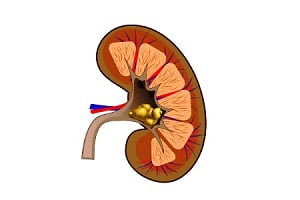
- Blood urea nitrogen (BUN) and creatinine is used to measure the functioning of kidney. Highly increased levels of urea indicate that stomach cancer has spread to the ureters or kidneys.
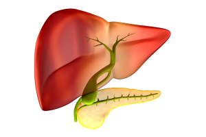
- Lactate acid dehydrogenase, alkaline phosphatase, transaminase and bilirubin are measured for proper functioning of liver. Their extreme high levels could indicate that cancer has spread to the liver.
Imaging tests for stomach cancer
X-ray
An X-ray produces a picture of the structures inside of the body by using small amount of radiations. It also helps in determining whether the stomach cancer has spread to the lungs or if there is any other serious lung diseases or heart diseases present.
Ultrasound for stomach cancer
Ultrasound mechanizes high-frequency sound waves to form pictures of structures in the body.
It is sometimes done along with endoscopy to study the spread of cancer which might have spread into the wall of the stomach and to the lymph nodes or surrounding tissues to the liver (detected in a liver ultrasound).
Ultrasound is also used to guide a needle for biopsy.
Computed tomography (CT or CAT) scan for diagnosing stomach cancer
A CT scan produces detailed images of soft tissues in the body. It mechanizes x-rays to make detailed 3-D cross-sectional images of the organs, tissues, bones and blood vessels using special X-ray equipment. A computer converts the images into detailed pictures. A CT scan is the most common and an effective way for staging advanced stomach cancer but not for finding the stomach tumors at an early stage.
A dye (contrast medium) may be given by mouth, injected into a vein (given intravenously) before a CT scan procedure to differentiate the affected part from the remaining tissues and allow capturing images better.
CT scan is used to:
- check the cancer spread to other tissues and organs, such as the lymph nodes or liver or to the ovary in women
- check for thickening of the stomach wall
Magnetic resonance imaging (MRI) scan
MRI scans produce detailed 3D cross sectional images of soft tissues such as organs, tissues, bones and blood vessels in the body.MRI scans use strong radio waves and powerful magnetic waves instead of x-rays.
An MRI is used for the detection and study of cancer in the stomach or cancer that has spread outside of the stomach.
Positron emission tomography (PET) or PET-CT scan
A slightly radioactive form of sugar called radiopharmaceuticals brings metabolic changes in the activity of body tissues. A special camera is then used to create pictures of areas of radioactivity in the body. Sometimes a PET scan may be done at the same time as a CT scan (called PET/CT scan) which brings areas that “light up” on the PET scan in more detail.
Laparoscopy for stomach cancer diagnosis
A laparoscope is used for the examination or for the removal of internal organs through several small incisions (surgical cuts) in the skin. Some very small tumors which cannot be detected through MRI and CT scan are found through laparoscopy. A thin flexible tube with a camera on its edge is inserted through a surgical cut to observe the surfaces of affected organs and nearby lymph nodes closely.
If there is no spread of the cancer, the abdomen is given a “wash” with saline (salt water), this is called peritoneal washing. The fluid is then removed to check if it contains cancer cells or not. If it does, the cancer has spread, even if the spread couldn’t be viewed through imaging.
Laparoscopy is used for staging stomach cancer which helps in preparing treatment plan for a patient.
The main objectives of a laparoscopy for stomach cancer are:
- observing the serosa (outside of the stomach) and nearby lymph nodes for cancer cells
- detecting cancer in other parts of the abdomen, such as the peritoneum or liver, which cannot be detected in a CT scan or an ultrasound
- performing biopsy (laparoscopic biopsy)
- removing fluid (peritoneal washings) from the abdomen to check for cancer cells
Stages of stomach cancer
The staging system most commonly used to stage a stomach cancer is the American Joint Committee on Cancer (AJCC) TNM system.
AJCC is based on 3 key pieces of information:
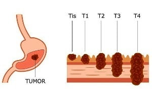
The extent (size) of the tumor (T): It defines the growth of cancer into the 5 layers of the stomach wall or its reach to nearby structures or organs.
The spread to nearby lymph nodes (N): The extension of cancer to nearby lymph nodes
The spread (metastasis) to distant sites (M): the cancer spread to distant lymph nodes or distant organs such as the liver or lungs
Stage 0 (Tis, N0, M0) stomach cancer
Tis : This stage is also known as carcinoma in situ (Tis). Sometimes, there are abnormal looking cells i.e. high grade dysplasia in the stomach lining. The tumor cells are only in the top layer of the mucosa (innermost layer of the stomach) and have not grown into the deeper layers of tissues such as the lamina propria
N0: The stomach cancer has not spread to nearby lymph nodes or distant sites.
M0: The stomach cancer has not spread to distant sites.
Stage 1 stomach cancer
1A (T1, N0, M0)
T1: The tumor has spread from the top layer of cells of the mucosa into the next below layers such as the lamina propria, the muscularis mucosa, or submucosa.
N0: It has not spread to nearby lymph nodes or distant sites.
M0: It has not spread to distant sites.
1B (T1 or T2, N1, M0)
T1: The cancer grows from the top layer of cells of the mucosa into the next layers below such as the lamina propria, the muscularis mucosa, or submucosa
Or,
T2: The cancer spreads into the muscularis propria layer.
N1: The cancer spreads to 1 to 2 nearby lymph nodes.
M0: It has not spread to distant sites (M0).
Stage 2 stomach cancer
2A (T1, N2, M0) OR (T2, N1, M0) OR (T3, N0,M0)
T1 AND N2: The cancer has grown from the top layer of cells of the mucosa into the next layers below such as the lamina propria, the muscularis mucosa, or submucosa (T1) AND it has spread to 3 to 6 nearby lymph nodes (N2).
OR,
T2 AND N1: The cancer grows into the muscularis propria layer (T2) AND spreads to 1 to 2 nearby lymph nodes (N1).
T3 AND N0: The cancer has grown into the subserosa layer (T3) AND has not spread to the nearby lymph nodes (N0).
M0: It has not spread to the distant sites.
2B (T1, N3a, M0) OR (T2, N2, M0) OR (T3, N1, M0) OR (T4a, N0, M0)
T1 and N3a: The cancer grows from the top layer of cells of the mucosa into the next layers below such as the lamina propria, the muscularis mucosa, or submucosa (T1) and it spreads to 7 to 15 nearby lymph nodes(N3a).
T2 AND N2: The cancer spreads into the muscularis propria layer (T2) AND it has spread to 3 to 6 nearby lymph nodes (N2).
T3 AND N1: The cancer grows into the subserosa layer (T3) AND it has spread to 1 to 2 nearby lymph nodes (N1).
T4a AND N0: The tumor has grown through the stomach wall into the serosa, but the cancer does not grow into any of the nearby organs or structures (T4a). It has not spread to the nearby lymph nodes (N0).
M0: The cancer has not spread to the distant sites.
STAGE 3 stomach cancer
3A (T2, N3a, M0) OR (T3, N2, M0) OR (T4a, N1, M0) OR (T4a, N2, M0) OR (T4b, N0, M0)
T2 AND N3a: The cancer has grown into the muscularis propria layer (T2) AND it has spread to 7 to 15 nearby lymph nodes (N3a)
OR,
T3 AND N2: The cancer grows into the subserosa layer (T3) AND it has spread to 3 to 6 nearby lymph nodes (N2).
OR,
T4a AND N1: The cancer has grown through the stomach wall into the serosa, but it has not grown into any of the nearby organs or structures (T4a). It has spread to 1 to 2 nearby lymph nodes (N1)
OR,
T4a AND N2: The cancer has grown through the stomach wall into the serosa, but it has not extended to any of the nearby organs or structures (T4a). It has spread to 3 to 6 nearby lymph nodes (N2)
OR,
T4b AND N0: The cancer has grown through the stomach wall and into nearby organs or structures (T4b). But it has not spread to the nearby lymph nodes (N0).
M0: In all cases, the cancer has not spread to the distant sites (M0).
3B (T1, N3b, M0) OR (T2, N3b, M0) OR (T3, N3a, M0) OR (T4a, N3a, M0) OR (T4b, N1, M0) OR (T4b, N2, M0)
T1 AND N3B: The cancer spreads from the top layer of cells of the mucosa into the next layers below such as the lamina propria, the muscularis mucosa, or submucosa (T1) AND it has spread to 16 or more nearby lymph nodes (N3b).
OR,
T2 AND N3b: The cancer is growing into the muscularis propria layer (T2) AND it has spread to 16 or more nearby lymph nodes (N3b).
OR,
T3 AND N3a: The cancer grows into the subserosa layer (T3) AND it has spread to 7 to 15 nearby lymph nodes (N3a).
OR,
T4a AND N3b: The cancer has grown through the stomach wall into the serosa, but it has not grown into any of the nearby organs or structures (T4a) AND it has spread to 7 to 15 nearby lymph nodes (N3a).
OR,
T4b AND N1: The cancer has grown through the stomach wall and into nearby organs or structures (T4b). It has spread to 1 to 2 nearby lymph nodes (N1).
T4b AND N2: The cancer has grown through the stomach wall and into nearby organs or structures (T4b). It has spread to 3 to 6 nearby lymph nodes (N2).
M0: Common in all, the cancer has not spread to the distant sites (M0).
3C (T3, N3b, M0 or T4a, N3b, M0 or T4b, N3a, M0 or T4b, N3b, M0)
T3 and N3b: The cancer is growing into the subserosa layer (T3) AND it has spread to 16 or more nearby lymph nodes (N3b).
OR,
T4a AND N3b: The cancer has grown through the stomach wall into the serosa, but it has not grown into any of the nearby organs or structures (T4a) AND it has spread to about 16 or more nearby lymph nodes (N3b).
T4b AND N3b: The cancer has grown through the stomach wall and into nearby organs or structures (T4b) AND it has spread to 7 to 15 nearby lymph nodes (N3a).
T4b AND N3b: The cancer has grown through the stomach wall and into the nearby organs or structures (T4b) AND it has spread to 16 or more nearby lymph nodes (N3b).
M0: In all cases, the cancer has not spread to the distant sites (M0).
Stage 4 (Any T, Any N, M1) stomach cancer
Any T, Any N AND M1: The cancer can grow into any layers (Any T) and might or might not have spread to the nearby lymph nodes (Any N). The cancer grows and spreads to the distant organs such as the liver, lungs, brain, or the peritoneum (the lining of the space around the digestive organs) (M1).
[Staging data inputs: American Cancer Society]Treatment of stomach cancer
Treatment for stomach cancer depends on several factors, including the severity of the cancer type and stage of the cancer, possible side effects, and an individual’s overall health and preferences.
Surgery for Stomach Cancer
Surgery is the complete removal of the cancer tumor from the affected part of the stomach and sometimes from the nearby lymph nodes, depending on the type and stage of stomach cancer. At stage 0, I, II, or III cancer, surgery along with other treatments is supposedly the only realistic option to cure the cancer, if the patient is healthy enough.
The different kinds of surgeries performed to treat the stomach cancer:
Endoscopic resection
Endoscopic mucosal resection is the preferred treatment option during an early stage stomach cancer from the stomach lining. The growth of tumor in the lymph nodes is also minor at this stage. An endoscope which is a long, flexible tube with a small video camera at its edge is made to pass down the throat and into the stomach.
Without any incision on the skin, the tumor is removed. A few surgical tools are passed with the endoscope to remove the affected part of the normal stomach wall around it.
Limited surgical resection is also an option which removes a section of the stomach wall by observing the tumor and the nearby areas through an endoscope to treat early stage stomach cancer.
Gastrectomy for stomach cancer treatment
Gastrectomy is the most common surgical treatment for stomach cancer.
Subtotal (partial) gastrectomy: Subtotal (partial) gastrectomy is often recommended if the cancer has started to grow only in the lower part of the stomach and very rarely it is done for cancers that are located only in the upper section of the stomach. Sometimes, only a part of the stomach is removed, and at times a part of the esophagus or the first part of the small intestine (the duodenum) is removed. The remaining section of the stomach is then re-attached after the surgery.
Total gastrectomy: If the cancer has spread throughout the stomach, total gastrectomy is done. It is more commonly done when the cancer begins in the upper part of the stomach, near the esophagus.
Total gastrectomy results in the removal of the entire stomach with the nearby lymph nodes, and omentum, and sometimes also involves the removal of the spleen and parts of the esophagus, intestines, pancreas, or other nearby organs.
The end of the esophagus is then attached to part of the small intestine which allows food to move down the intestinal tract. Consumption of food becomes a problem after this surgery.
Lymph node removal
Lymph node dissection is mostly done along with a gastrectomy to remove lymph nodes around the stomach. Removal of a minimum of 15 lymph nodes is called a D1-lymphadenectomy when a gastrectomy is done. When more than lymph nodes near the cancer are removed, it is called a D2 lymphadenectomy.
Other surgical methods for removing stomach tumors
- Laparoscopic staging and exam is conducted if the cancer grows from the stomach to other parts of the abdomen, such as the liver or pancreas.
- Feeding tube placement: Placing a feeding tube through the skin of the abdomen and then into the distal part of the stomach with a minor operation, is known as a gastrostomy tube surgery or G tube or jejunostomy tube or J tube (into the small intestine). It is done to provide food and nutrition to patients who have problems in consuming food after the surgery.
- When the cancer has spread widely in the body, surgery is preferred because it prevents the bleeding from the tumor and also prevents the stomach from being blocked by the growth of the tumor. This is called palliative surgery and it is done to prevent the symptoms from occurring. Palliative surgeries can be gastric bypass, endoscopic tumor ablation, stent placement, etc.
Possible complications and side effects of stomach cancer surgery
- Side effects of surgery include bleeding from the surgery, blood clots, and damage to the nearby organs during the operation.
- The new connections which are made between the ends of the stomach or esophagus and small intestine may leak.
- Other side effects include nausea, heartburn, abdominal pain, and diarrhea, particularly after eating.
- Changes in diet will be needed after a partial or total gastrectomy.
Chemotherapy for Stomach Cancer
Anti-cancer drugs are injected into the veins or given orally as pills which enter the bloodstream and metastasize in all areas of the body. This is known as chemotherapy. Chemo-treatment is useful for any cancer that has spread to multiple organs.
Chemo can be used in different ways to help treat stomach cancer:
- Neoadjuvant treatment: Chemotherapy is given before surgery for stomach cancer which helps the tumor to shrink. It also helps to keep the cancer from coming back and thus increases the lifespan of a patient.
- Adjuvant treatment: Chemo is given after the surgery to remove the cancer that is left behind. The goal of adjuvant chemo is to kill any cancer cells that might have been left behind. This restrains the cancer from coming back. Often, for stomach cancer, chemo is given with radiation therapy after the surgery. This is called chemoradiation therapy.
A number of chemo drugs given during chemotherapy for stomach cancer are:
- 5-FU (fluorouracil), often given along with leucovorin (folinic acid)
- Oxaliplatin (Eloxatin®)
- Cisplatin
- Irinotecan (Camptosar®)
- Epirubicin (Ellence®)
- Docetaxel (Taxotere®)
- Paclitaxel (Taxol®)
- Carboplatin
- Capecitabine (Xeloda®)
Some common drug combinations used when surgery is planned include:
- In most chemotherapy treatments for stomach cancer, a combination of at least 2 drugs, Cisplatin (Platinol) and Fluorouracil (5-FU, Adrucil) is given.
- ECF (epirubicin, cisplatin, and 5-FU), given before and after the surgery.
- Docetaxel or paclitaxel plus either 5-FU or capecitabine, used during chemoradiation as treatment before the surgery.
- Cisplatin plus either 5-FU or capecitabine, combined with radiation therapy before the surgery.
- Paclitaxel and carboplatin, combined with radiation as treatment before the surgery.
For the advanced treatment of stomach cancer, ECF may also be used, but other combinations may also be helpful. Some of these include:
- DCF (docetaxel, cisplatin and 5-FU)
- Irinotecan plus cisplatin
- Irinotecan plus 5-FU or capecitabine
- Oxaliplatin plus 5-FU or capecitabine
Chemo may be given as the primary treatment for stomach cancer that has spread (metastasized) to distant organs. It may help shrink the cancer or slow its growth, which can relieve symptoms for some patients and help them live longer.
Side effects of chemotherapy
- Nausea and vomiting
- Hair loss
- Fatigue and shortness of breath (shortage of red blood cells)
- Loss of appetite
- Diarrhea
- Mouth sores
- Increased risk of infection (shortage of white blood cells)
- Bleeding or bruising after minor cuts or injuries (from a shortage of platelets)
- Heart damage: Doxorubicin, epirubicin, and some other drugs cause permanent heart damage if they are used for a long time or in high doses.
- Hand-foot syndrome: During treatment with capecitabine or 5-FU (given as an infusion), the beginning of redness in hands and feet progresses to pain and sensitivity in the palms and soles. Blistering or skin peeling sometimes occur in worse cases which leads to open, painful sores.
- Cisplatin, oxaliplatin, docetaxel, and paclitaxel can cause great damage to the nerves outside the brain and spinal cord. Some rare symptoms may also occur such as pain, burning or tingling sensations, sensitivity to cold or heat, difficulty in eating and drinking or weakness(mainly in the hands and feet).
Targeted Therapies for Stomach Cancer
Targeted therapy limits the growth of cancer cells in the stomach. The therapy targets cancer specific genes, proteins, or the tissue environment which contributes to the cancer cell spread and survival. It does not damage healthy cells largely.
Research studies have found some specific molecular targets and new treatment therapies, such as:
- HER2-targeted therapy: A protein called human epidermal growth factor receptor 2 (HER2) is sometimes excessively produced by some tumors which leads to cancer growth. This type of cancer is called HER2-positive cancer. Trastuzumab (Herceptin) plus chemotherapy is an option for treating later-stage HER2-positive stomach cancers.
- Anti-angiogenesis therapy: Anti-angiogenesis therapy is a type of targeted therapy which focuses on stopping the angiogenesis. Angiogenesis is the process of making new blood vessels which deliver nutrients needed for the growth and expansion of the tumor. The aim of anti-angiogenesis therapy is to “starve” the tumor.
Some new targeted drugs have been developed for the treatment of stomach cancer:
Trastuzumab
Trastuzumab or Herceptin targets the HER2 protein. It is a monoclonal antibody, a man-made version of a very specific immune system protein. This drug only works if the cancer cells have excess of HER2.
Side effects of trastuzumab are relatively mild. Common side effects are fever and chills, weakness, nausea, vomiting, cough, diarrhea, and headache.
The chances of any kind of heart damage increases if trastuzumab is combined with certain chemo drugs called anthracyclines, such as epirubicin (Ellence) or doxorubicin (Adriamycin).
Ramucirumab
Ramucirumab or Cyramza is used during the treatment of advanced stage stomach cancer, when other drugs do not give a positive output.
Ramucirumab is a monoclonal antibody that binds to a receptor or to cell surface proteins and signals the body for producing more blood vessels. This lowers down or stops the growth and spread of cancer.
The most common side effects are high blood pressure, headache, and diarrhea. Some serious possible side effects which are not as frequently noted are:
- blood clots
- severe bleeding
- holes forming in the stomach or intestines (perforations)
- problems with wound healing
Some other drugs are: Imatinib (Gleevec), Sunitinib (Sutent) and Regorafenib (Stivarga) for a rare form of stomach cancer called gastrointestinal stromal tumor.
Immunotherapy for Stomach Cancer
Immunotherapy, which is also known as biological therapy, boosts the body’s natural defense mechanism to fight cancer. Medicines used in immunotherapy are either produced by the body or in a laboratory. These medicines improve, target, and restore the function of immune system. Immunotherapy is an active and prominent area of research for the stomach cancer.
Immune checkpoint inhibitors: Some molecules on immune cells are required to be turned on (or off) to start an immune response known as the checkpoints. Cancer cells sometimes use checkpoints to save themselves from being attacked by the immune system. Therefore, new drugs that target these checkpoints are used for cancer treatments. For example, Pembrolizumab (Keytruda) targets PD-1 which is a protein on T cells that normally helps these immune cells from attacking other cells in the body.
Pembrolizumab boosts the immune response against cancer cells. This can shrink some tumors or slow their growth.
Possible side effects of immunotherapy
- Feeling tired or weak
- Fever and chills
- Cough
- Nausea
- Itching and rashes
- Loss of appetite
- Muscle or joint pain
- Shortness of breath
- Constipation or diarrhea
- Flushing of the face
- Feeling dizzy, wheezing, and trouble breathing
- Autoimmune reactions: Sometimes the immune system start attacking other parts of the body, which cause serious or even life-threatening problems in the lungs, intestines, liver, endocrine glands, kidneys, skin, or other organs.
Radiation Therapy for Stomach Cancer
High-energy X-rays or other particles are used to destroy cancer cells in a specific body area through radiation therapy. A radiation therapy schedule consists of a specific number of set treatments given over a set period of time.
Radiations are generally used along with chemotherapy (chemo) to shrink the tumor in size and to make the surgery easier. Small remains of the cancer can still stay in the stomach which may not be visible during the surgery. These are destroyed by radiation therapy. Radiation therapy is often used to decrease the cancer growth and ease the symptoms of advanced stomach cancer such as pain, bleeding, and eating problems.
External beam radiation therapy (EBRT)
EBRT uses radiations which focus on the cancer from a machine outside the body. Three-dimensional conformal radiation therapy (3D-CRT) and intensity modulated radiation therapy (IMRT) are used often as special techniques which focus the radiations on the affected part to kill cancer cells from different angles and also limit the damage to nearby normal tissues. The beams are aimed on certain parts of the body and other parts are shielded from the radiation during treatment.
Side effects from radiation therapy for stomach cancer
Radiations damage the nearby organs which lead to problems such as heart or lung damage, or even an increased risk of another cancer later. Therefore, only required and limited dosage of radiation is given to the patient.
Other side effects of radiation therapy are:
- Skin problems, such as redness, blistering, peeling, in the area exposed to radiations
- Low blood cell counts
- Fatigue
- Nausea and vomiting
- Diarrhea
Treatment Choices by Type and Stage of Stomach Cancer
A stomach cancer grows and spreads in different ways. It can grow through the wall of the stomach and advance to the nearby organs and also to the lymph vessels and nearby lymph nodes.
The stomach cancer cells can travel through the bloodstream and metastasize to organs such as the liver, lungs, and bones, which are hard to treat.
Treatment of Stage 0 stomach cancer
Stomach cancers are found only in the inner lining of the stomach and not into deeper layers. Stomach cancer can be treated completely by surgery either through total or subtotal gastrectomy. No chemotherapy or radiation therapy is required at this stage.
Endoscopic resection surgery procedure is used to remove small size cancer tumors through an endoscope.
Treatment of Stage 1 stomach cancer
Stage 1a: Stage 1a stomach cancer is removed by total or subtotal gastrectomy. Nearby lymph nodes are also removed.
Stage 1b: At stage 1b, stomach cancer is removed by surgery (total or subtotal gastrectomy). If the cancer has also spread to the lymph nodes, treatment with either chemo-radiation or only chemo therapy is recommended.
Treatment of Stage 2 stomach cancer
The primary treatment for stomach cancer at this stage is surgery which involves removal of all or part of the stomach, the omentum, and the nearby lymph nodes. Chemotherapy or chemoradiation therapy is given before the surgery to reduce the size of the cancer which makes it easier to be removed.
Treatment of Stage 3 stomach cancer
Surgery can be beneficial in some cases but chemotherapy and radiation therapy are a must to treat the cancer in the stomach and to the lymph nodes, and sometimes to the nearby organs. The cancer can remain if the chemo or chemo-radiation therapy is not given both before and after chemotherapy.
Treatment of Stage 4 stomach cancer
At this stage, the stomach cancer has spread to distant organs of which the cure is usually not possible. The treatment only helps in keeping the cancer under control and helps in relieving the symptoms.
The treatment includes surgery, such as a gastric bypass or even a subtotal gastrectomy (in some cases), chemo and/or radiation therapy (to shrink the cancer tumor), targeted therapy and immunotherapy to treat cancers at advanced stage.
After the Cancer Treatment, Living as a Stomach Cancer Survivor
There are strong chances of cancer to reoccur even after successful treatment. Even this thought of cancer recurrence makes any cancer survivor stressed. In some cases, cancer might never go completely. These are certain mental challenges a cancer patient has to face in his or her life.
In case of a stomach cancer, food consumption also reduces and therefore the diet of a person also changes according to cancer stages.
In some patients with stomach cancer, problems such as nausea, diarrhea, sweating, and flushing become common after eating. This is called dumping syndrome.
When parts or complete stomach is removed, the food that is consumed is swallowed quickly and is passed into the intestine fast, which may cause discomfort and other digestion-related symptoms after eating. These symptoms get better over time, though.
Sometimes, nutritional supplements are provided with the help of a feeding tube, usually called a jejunostomy tube (or J-tube), which is put into the small intestine by making a small hole in the skin over the abdomen during a minor operation. J-tube allows liquid nutrition to flow directly into the small intestine to prevent loss in weight and improve nutrition. Sometimes, though rarely, the tube is placed into the lower part of the stomach. This is known as a gastrostomy tube or G-tube.
Medical records are to be maintained by patients and their caretakers for the treatment of stomach cancer at all stages and also for future use if the cancer reoccurs. Health insurance is also necessary because the treatment of stomach cancer is expensive.
Spread of the stomach cancer: What are the common sites where stomach cancer can spread to?
If the stomach cancer grows, it spreads to the following areas easily:
- lymph nodes in and around the abdomen or above the left collarbone (Virchow node)
- small intestine
- spleen
- mesentery (folds of tissue that hold the abdominal organs in place)
- omentum
- pancreas
- colon
- liver
- abdominal wall
- diaphragm
- esophagus
- adrenal glands
- lung
- bone
- pelvic area around the rectum (Blumer shelf)
- area around the belly button
- skin
- ovaries (Krukenberg tumour)
- uterus
- brain
How can you reduce the risk of recurrence of stomach cancer?
Exercising, eating a certain type of diet, and taking nutritional supplements can only help a patient reduce the risk of cancer. But, it cannot cure the cancer or prevent the cancer from returning completely.
The use of tobacco is clearly a risk factor for stomach cancer. Reducing its consumption can reduce the risk. This can also help in improving the appetite and can also reduce the chance of developing other types of cancer.
Re-occurrence of stomach cancer
Recurrent cancer or reoccurrence means that the cancer has come back after successful treatment. It might be in the same area from where it had started in the first case or at a different location.
If the stomach cancer returns, there is another round of tests to diagnose the spread and its recurrence. The cancer might spread in the stomach or in other parts of the body such as the liver, lymph nodes etc.
Often, the stomach cancer cannot be detected at early stages and therefore it becomes difficult to treat the patient. It is also sometimes unfortunate that people with stomach cancer cannot survive the disease.
