How to Read Dental X Rays or Radiographs for Cavities?
- Updated on: Jul 1, 2024
- 3 min Read
- Published on Apr 19, 2021
Understanding Dental X-Rays
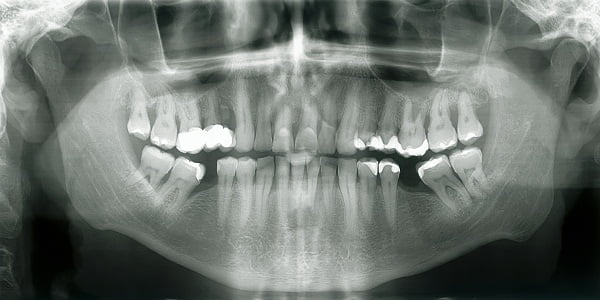
Dental X-rays are an essential part of any oral care diagnosis. They are quite commonly used to make a diagnosis of dental cavities or other dental problems. If you have ever visited a dentist, you probably might know about them and how they are recorded. It is good to learn more about them and know about reading them.
A dentist or a radiologist will cover you with a heavy iron apron to protect your body from the radiation. He will then insert a small apparatus, made of plastic, into your mouth and ask you to bite down on it to hold the X-ray film in place. The technician will then take an X-ray picture of the targeted area. This process is pain-free.
How do Cavities appear under X-ray?
Cavities manifest as dark blemishes on a tooth when viewed through an x-ray. They initially form in the enamel, the hardest and outermost layer of the tooth. On an x-ray, enamel appears as the brightest shade. Over time, the cavity can progress deeper into the tooth, reaching the dentin. Dentin is softer than enamel, making it more susceptible to decay, and it appears as a darker shade on the x-ray compared to enamel. Notably, dental x-rays are crucial in identifying cavities that form between adjacent teeth, areas that are typically hidden from direct view and would go unnoticed to the unaided eye.
How to read or interpret an X-ray?
This article can’t make you an expert but this page outlines how dentists read x-rays.
Here are some tips for you to read dental radiographs. You will however require a dentist to interpret them perfectly and to diagnose the condition and understand the severity of dental cavities if they exist.
What do black and white colors indicate on a dental X-ray?
- Soft objects appear black on x-ray
- Solid objects appear white on x-ray
What does grey/black color indicate on x-ray?
It can indicate any of these:
- Decay of tooth
- Abscess
- Pump (nerves or blood vessels)
- Gums in between the teeth spaces
What does white/cream color indicate on x-ray?
White/cream color indicates:
- Enamel
- Dentine as creamy white layer
- Metal fillings and crowns appear white
How does bone look like on the X-ray?
Bone appears as mottled grey and white in color. It has a white line at its margin around the teeth.
Detection of tooth decay (cavity or caries) on a dental X-ray
When looking for cavities, a dentist looks for signs on the radiograph (x-ray) that indicate a change in the density of any tooth’s enamel and/or dentin.
Tooth decay is a region of tooth de-mineralization (region with reduced minerals). Sometimes it is even an outright hole or pit. These locations where demineralization has happened or pits and holes have formed appear as a darkened area on the x-ray.
This is because the portion of the tooth that has decayed is less “hard” (less dense) and therefore the x-rays penetrate the portion of the tooth more easily and expose the film to make the area on the film look darker.
X-rays show tooth decay starting at the contact point between your teeth. This is not visible to the dentist with naked eyes. If dental cavities are discovered early, this can prevent the decay from spreading deeper into the tooth. Read about prevention of tooth decay (dental cavities): What Can I Do to Keep Cavities from Forming?
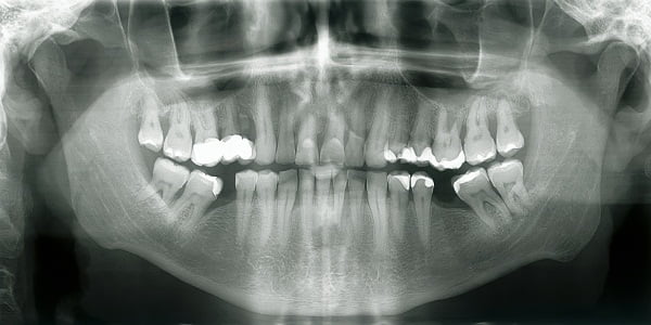
Image Credit: Drchristophermorris.com
The red arrow on this x-ray shows tooth decay (the dark area) between the teeth in a patient.
