


Browsing: Achondroplasia Graphics
Comprehensive Information, Resources, and Support on Achondroplasia
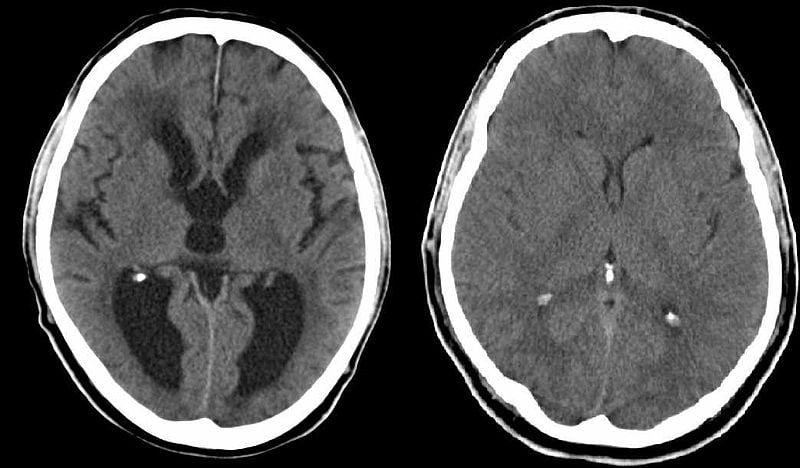
Hydrocephalus can be diagnosed through a CT scan, an MRI, or an ultrasound (in infants). These scans help in showing the build-up of cerebrospinal fluid on the brain and the increased pressure, as well as highlighting any structural defects that may be causing the problem. CT scans for hydrocephalus are used only during emergency examination. [Image used under Creative Commons License from Wikimedia]
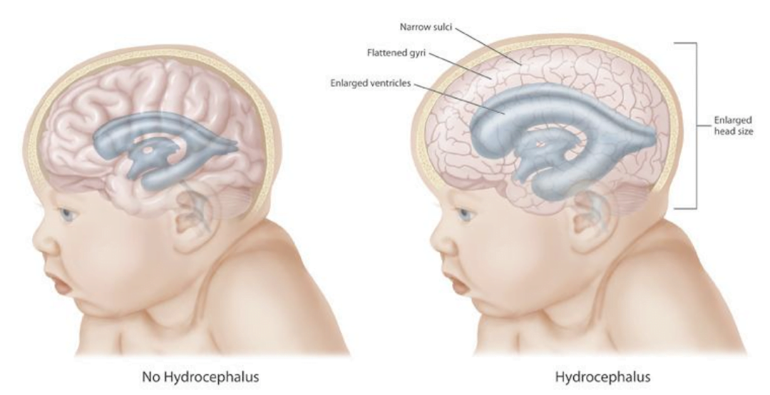
Hydrocephalus is a condition which can be explained as “water in the brain”. It commonly occurs in children suffering with achondroplasia or dwarfism. In this condition, the ventricles enlarge due to which the head size increases. The excess fluid in the brain increases the size of the ventricles or the cavities which puts pressure on the brain. This excess fluid is called cerebrospinal fluid (CSF). [Image used under Creative Commons License from Wikimedia]
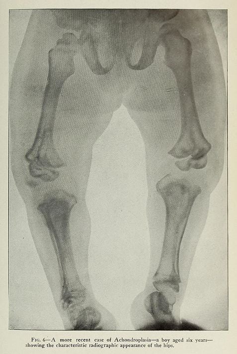
The picture depicts an X-ray diagram of a six year boy showing the characteristic radiographic appearance of the hips. In achondroplasia, a child’s arms and legs are short in proportion to body length. Radiographic imaging or X-ray scan of the infant may also be used to diagnose achondroplasia as the long bones show changes from the x-ray scans of non-affected infants. X-ray scans can also help in detecting shortened spinal pedicles distally with a decreased interpedicular distance in the inferior lumbar spine compared with the superior lumbar spine. [Image used under Creative Commons License from Wikimedia]
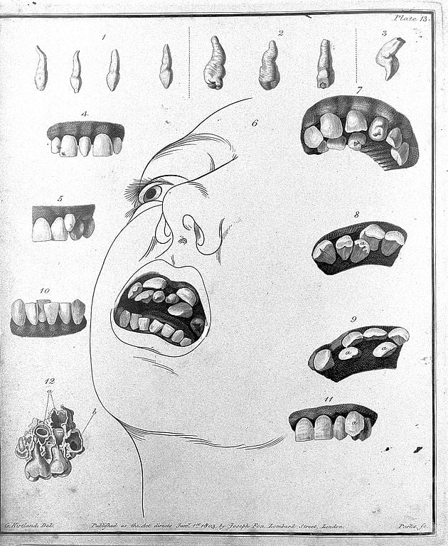
Teeth crowding or misalignment of teeth or crooked teeth is one of the symptoms associated with achondroplasia. Children suffering from achondroplasia experience bone deformities due to which the growing teeth also emerge as misaligned. In most cases, crooked teeth are due to shorter jaw. Panoramic radiography is used to detect the actual position of the teeth placed in the jaw. [Image used under Creative Commons License from Wikimedia]
ADVERTISEMENT