Advancements in Early Detection Technologies: A Ray of Hope in Breast Cancer Prevention
- Updated on: Mar 14, 2024
- 6 min Read
- Published on Mar 14, 2024
Breast cancer is a daunting disease that affects millions of women worldwide. However, with advances in medical science, the outcomes are significantly improved with early detection. Thanks to advancements in early detection technologies, there is now a ray of hope in the battle against this formidable foe. The evolution of these technologies has revolutionized breast cancer screening, making it faster, more accurate, and ultimately more effective in saving lives.
3D Mammography: A New Dimension in Breast Cancer Screening
Over the years, early detection technologies for breast cancer have come a long way in helping doctors diagnose the disease at its earliest stages. Mammograms have been the gold standard for screening, but they had their limitations. However, recent breakthroughs have paved the way for more innovative methods, ensuring that no stone is left unturned in the fight against breast cancer.
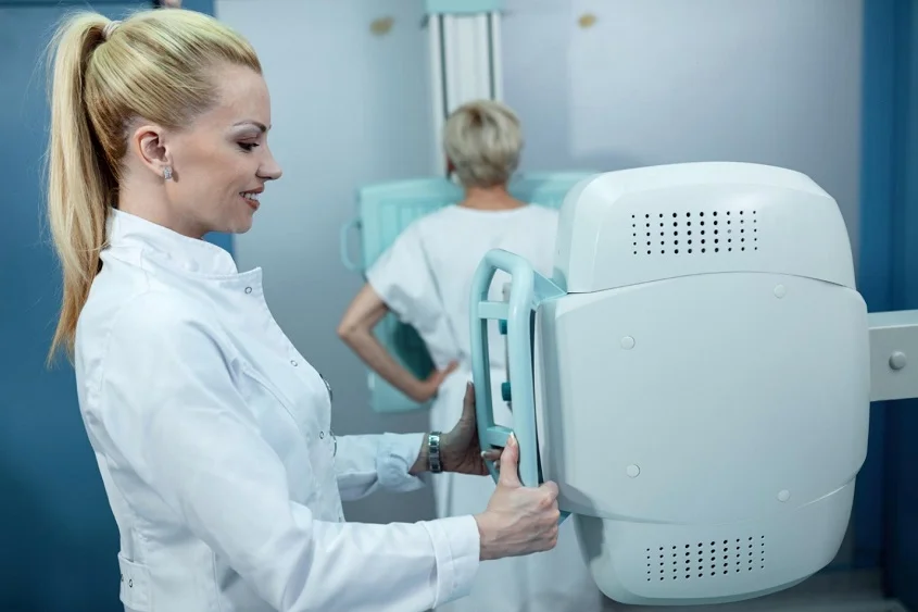
One of the most significant advancements in recent years has been the introduction of 3D mammography, also known as digital breast tomosynthesis. This cutting-edge technology allows radiologists to examine multiple images of the breast in greater detail, making it easier to spot even the tiniest of abnormalities. As a result, 3D mammography has been shown to improve cancer detection rates while reducing the number of false positives.

Traditional 2D mammograms provide a flat image of the breast tissue, which can sometimes overlap and create confusion for radiologists, specially with dense breast tissue. In contrast, 3D mammography captures multiple images of the breast from different angles, creating a three-dimensional view that helps doctors navigate through the layers of tissue more effectively. This improved clarity and precision can lead to earlier detection of breast cancer, ultimately saving lives.
Furthermore, the advanced imaging capabilities of 3D mammography make it particularly beneficial for women with dense breast tissue. Dense breasts have more glandular and fibrous tissue than fatty tissue, which can make it challenging to detect abnormalities on 2D mammograms. With 3D mammography, radiologists can examine the breast tissue layer by layer, making it easier to identify potential tumors hidden within dense areas. This increased accuracy can provide peace of mind for women with dense breasts, knowing that their screenings are more thorough and reliable.
Beyond Mammograms: Ultrasound and MRI Innovations
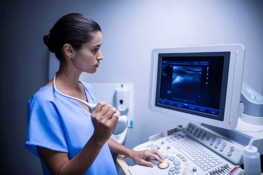
In addition to 3D mammography, ultrasound and magnetic resonance imaging (MRI) have emerged as valuable tools in breast cancer screening. Ultrasound utilizes sound waves to create detailed images of the breast, providing additional information that can help identify suspicious areas that may be missed by mammography alone. Likewise, MRI scans offer a more comprehensive view, particularly for high-risk patients, enabling early detection and intervention.
Ultrasound technology has advanced significantly in recent years, with the development of techniques such as elastography and contrast-enhanced ultrasound. Elastography measures tissue stiffness, aiding in the differentiation between benign and malignant lesions. Contrast-enhanced ultrasound involves injecting a contrast agent to enhance the visibility of blood flow, helping to characterize tumors more accurately. These advancements have improved the accuracy of ultrasound in detecting breast abnormalities, leading to more precise diagnoses and treatment planning.
MRI technology, on the other hand, has seen improvements in image resolution and contrast enhancement. Dynamic contrast-enhanced MRI is a technique that involves injecting a contrast agent to highlight areas of increased blood flow, which is often indicative of cancerous growth. Additionally, diffusion-weighted imaging can provide information about the cellular density of tissues, aiding in the characterization of lesions. With these advancements, MRI has become an indispensable tool in the early detection and monitoring of breast cancer, especially in cases where mammography results are inconclusive.
The Rise of Contrast-Enhanced Mammography (CEM) and Molecular Breast Imaging (MBI)
Continuing on the path of innovation, contrast-enhanced mammography (CEM) and molecular breast imaging (MBI) have gained traction in recent years. CEM utilizes a contrast agent to enhance the visibility of abnormalities, improving the accuracy of mammography even further. On the other hand, MBI employs a radioactive tracer that specifically targets breast cancer cells, providing a highly sensitive method of detection, particularly for women with dense breast tissue.
Contrast-enhanced mammography (CEM) represents a significant advancement in breast imaging technology. By introducing a contrast agent into the body, CEM is able to highlight areas of concern that may have been difficult to detect with traditional mammography alone. This enhanced visibility can lead to earlier detection of breast cancer, ultimately improving patient outcomes and survival rates.

Molecular breast imaging (MBI) offers a unique approach to detecting breast cancer at a molecular level. The use of a radioactive tracer allows MBI to identify even small clusters of cancer cells, making it a valuable tool for screening high-risk populations. Additionally, MBI is particularly beneficial for women with dense breast tissue, where traditional mammography may be less effective. By targeting cancer cells with precision, MBI helps reduce false positives and unnecessary biopsies, providing peace of mind for patients and healthcare providers alike.
Promising Biomarkers and Genetic Testing for Early Breast Cancer Detection
Advancements in genetics have opened up a whole new world of possibilities for early detection. Researchers have identified various biomarkers that can indicate the presence of breast cancer. Additionally, genetic testing can provide insights into an individual’s susceptibility to the disease, helping doctors tailor personalized screening strategies and intervention plans.
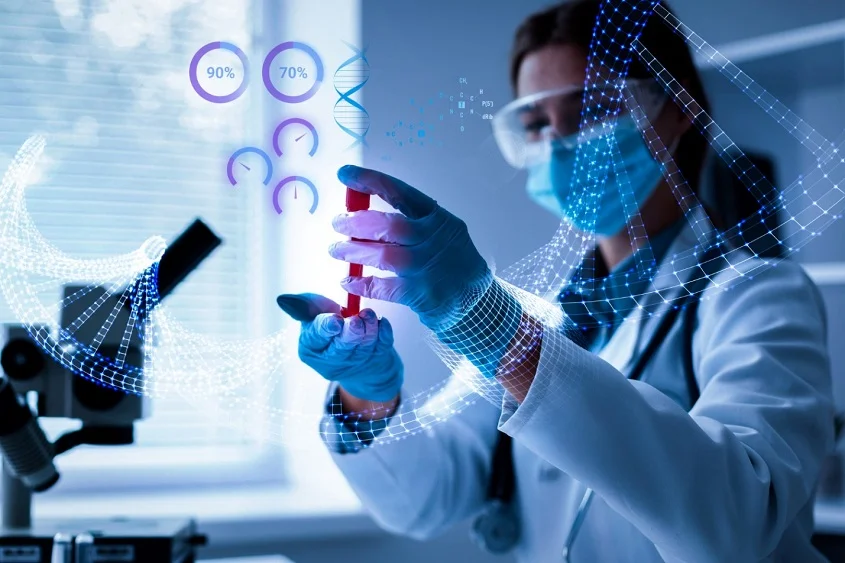
Moreover, researchers are exploring the potential of liquid biopsy as a minimally-invasive method for detecting breast cancer at an early stage. This cutting-edge technique involves analyzing circulating tumor cells and cell-free DNA in the blood to identify genetic mutations associated with breast cancer. Liquid biopsies can identify specific genetic mutations within the cancer DNA circulating in the blood. This information can help oncologists select targeted therapies that are most likely to be effective against the particular characteristics of a patient’s cancer.
Liquid biopsy shows great promise in not only detecting breast cancer early but also monitoring treatment response and detecting recurrence, revolutionizing the way breast cancer is diagnosed and managed. After treatment, liquid biopsies can be used to detect minimal residual disease or early signs of breast cancer recurrence, often before they are apparent on imaging tests or cause symptoms. This early detection can allow for timely interventions to address the recurrence.
The Role of Artificial Intelligence in Enhancing Early Detection of Breast Cancer
Artificial intelligence (AI) is revolutionizing many aspects of healthcare, and breast cancer detection is no exception. By analyzing vast amounts of imaging data, AI algorithms can quickly and accurately identify patterns and anomalies that may be indicative of breast cancer. This technology has the potential to assist radiologists in detecting cancer at its earliest stages, improving survival rates and reducing unnecessary invasive procedures.
Personalizing Prevention: Genetic Testing and Digital Health
In an era of personalized medicine, genetic testing and digital health tools have emerged as integral components of breast cancer prevention. Genetic testing can identify individuals at high risk for breast cancer, enabling them to take proactive measures such as increased surveillance or preventive surgeries. Simultaneously, digital health applications provide resources and support for patients, empowering them to take control of their breast health.
Empowering Patients through Education on Early Detection Methods
Empowering patients with knowledge about early detection methods is crucial in the fight against breast cancer. By educating women about the latest advancements in screening technologies, healthcare providers can encourage regular screenings, raise awareness about breast self-examinations, and emphasize the importance of seeking medical attention if any concerning symptoms arise. Through education, we can enable women to be proactive advocates for their own breast health.
For example, breast tissue density is something that every woman should be aware of. The chances of a missed diagnosis is highest in these women, and hence the women, armed with this knowledge, should advocate for themselves and request a 3D mammogram or other advanced screening modalities. Even they should also aware about the steps to take after cancer diagnosis.
Challenges and Opportunities in Implementing Early Detection Technologies
While advancements in early detection technologies offer tremendous promise, there are challenges that need to be addressed to ensure their widespread implementation. Access to these technologies, particularly in underserved communities, remains a concern. Furthermore, healthcare providers must stay abreast of the latest developments and receive specialized training to effectively utilize these cutting-edge tools.
The Future of Breast Cancer Screening
The journey towards conquering breast cancer continues, with new developments on the horizon. Researchers are exploring novel technologies, such as liquid biopsies and breathalyzer tests, which have the potential to revolutionize the field of early detection further. As the battle against breast cancer rages on, we can find hope in the continuous advancements in early detection technologies, providing a ray of light for a future free from the fear of this devastating disease.
