All About Leukemia (Blood Cells Cancer)
- Updated on: Jul 8, 2024
- 16 min Read
By
- Published on Oct 3, 2019
Overview and Definition: What is Leukemia?
Leukaemia is the general name given to a group of cancers that develop in the blood forming tissues and blood cells including the bone marrow and the lymphatic system. Leukaemia starts in developing blood cells that have undergone a malignant change.
Blood cells found in your body are broadly categorized as white blood cells (WBCs), red blood cells (RBCs) and platelets. Malignant blood cells multiply in an uncontrolled way and do not mature properly, leaving them unable to function as they should.
Every day, your body makes billions of new blood cells in the bone marrow especially the red blood cells. But during leukemia your body makes white cells more than it’s needed.
Leukemia is generally referred to the cancer of white blood cells. There are two main types of white blood cells in your body: lymphoid cells and myeloid cells. Leukemia can happen in either of them.
WBCs are a very important member of your immune system and mainly help to fight external or internal infection of the body. They also protect your body from invasion by bacteria, viruses, and fungi and other foreign substances. Your body starts producing abnormal WBCs in leukemia, which do not function properly. They increase in number by dividing quickly which eventually crowd out the normal cells of the blood.
Stems cells of the blood is the precursor for production of different kinds of blood cells. Stem cells develop into immature blood cells known as blasts and these blasts ultimately turn into WBC, RBC and platelets. Overproduction of these blast cells causes leukaemia. In leukaemia, these blast cells develop abnormally and don’t convert into mature blood cells. With passage of time, these blast cells increase in number and crowd out the normal blood cells so that they can’t do their jobs well. When the diagnosis of leukemia is confirmed, these blast cells may be called as leukemia cells.
There are many types of leukemia based on the type of blast cells in which they develop. Some of them are more common in children while other forms of leukemia occur mostly in adults. Leukemia is misunderstood as a condition more prevalent in children, but it actually affects adults more. Generally, leukemia is more common in men than in women. White population is more susceptible to leukemia than African-Americans.
Depending on the type of leukemia and other factors, the treatment for leukemia can be complex but there are strategies and resources that can help to make your treatment successful. There is no known mechanism to prevent leukemia.
How is leukemia classified? What are the different types of leukemia?
Leukemia is mainly classified on the basis of the type of blast cells from which they originate and the speed of their progression.
On the basis of cell type involved, leukemia is classified as:
- Lymphocytic leukemia
- Myelogenous leukemia
On the basis of their speed of progression, leukemia is classified as:
- Acute leukemia
- Chronic leukemia
Read more about various types of leukemia.
What are the signs and symptoms of leukemia?
Signs and symptoms of leukemia depend mainly on the type of leukemia, but common signs and symptoms of leukemia are:
- Fever or chills
- Persistent fatigue, weakness that don’t go away with rest
- Frequent or severe infections
- Unintentional weight loss
- Paleness
- Rapid heartbeat
- Shortness of breath
- Bleeding gums
- Swelling in the testicles
- Vision problems
- Sores in the eye
- Sore throat
- Chloroma – a collection of leukemia cells, or blasts, under the skin or in other parts of the body
- Leukemia cutis – appears as sores or as patches of any size that are usually pink or tan in colour
- Swollen lymph nodes, enlarged liver or spleen
- Easy bleeding or bruising
- Recurrent nosebleeds
- Tiny red spots in your skin (petechiae)
- Excessive sweating, especially at night
- Bone pain or tenderness
What causes leukemia? How do you get leukemia?
There is no exact known cause of leukemia and it is thought to develop because of combined effect of genetic and environmental factors. As with any cancer, leukemia happens when the DNA of immature blood cells, mainly white cells, becomes damaged. This damage of the DNA causes the blood cells to grow and divide continuously without dying, so that there are too many of such cells in the body. In normal conditions, healthy blood cells die after some time and are replaced by new cells, which are produced in the bone marrow. But if you have leukemia, abnormal blood cells do not die at their right rate. They accumulate and occupy more space leaving a little space for other normal blood cells to grow.
As the number of leukemia cells increases, they crowd out the space in the blood which in turn stops the healthy white blood cells from growing and functioning normally.
What are the risk factors for developing leukemia? Who can develop leukemia?
Some lifestyle habits and environmental factors are supposed to increase the risk of leukemia. However, these risk factors do not always cause the cancer. Many patients diagnosed with leukemia are found to be devoid of any associated risk factor. Some of the risk factors for leukemia include:
Previous cancer treatment
Cancer therapy given in the form of chemotherapy and radiation therapy for the treatment of some other cancers or medical conditions in the past may increase the risk of certain types of leukemia.
Genetic disorders
Genetic abnormalities or genetic mutations seem to play a role in the development of many types of cancers including leukemia. These genetic changes are known as inherited cancer syndromes and they can occur in both – children and adults. There are certain genetic disorders, such as Down syndrome, that are associated with an increased risk of leukemia. Other genetic disorders which can be considered a potential risk factor for leukemia are:
- Fanconi anemia
- Ataxia-telangiectasia
- Bloom syndrome
Exposure to certain chemicals
Exposure to certain chemicals, such as benzene and formaldehyde are linked to an increased risk of some kinds of leukemia. Benzene is present in gasoline and people working in chemical industry may breathe in the benzene environment. People working as embalmers have higher risk of developing leukemia because they tend to have longer contact with formaldehyde.
Smoking
Cigarette smoking increases the risk of acute myelogenous leukemia (AML).
Family history of leukemia
Your risk of leukemia increases if any member in your family has been diagnosed with leukemia. For example, in case of identical twins, if one gets a certain type of leukemia, there is a 20% chance that the other twin will have it within a year.
What are the complications of leukemia?
Most of the problems related to leukemia are due to depletion of the normal blood cells as well as the side effects of various treatments. These may include such as frequent infections, bleeding, and graft-versus-host disease (GVHD) in recipients of stem cell transplants. Weight loss and anaemia are two major visible complications of leukemia and its treatment. Relapse or progression of the disease after its remission with successful treatment is also a major concern in people with leukemia.
There are other complications of leukemia which are specific to the type of leukemia. For example, in 3% to 5% of cases of chronic lymphocytic leukemia (CLL), the cells transform into an aggressive lymphoma by changing their characteristics which is known as a Richter transformation. One of the potent complications of CLL is autoimmune hemolytic anemia, where your own body attacks and destroys the red blood cells. People having CLL are at greater risk of second cancers and other blood disorders and blood cancers.
Tumor lysis syndrome is seen in some cases of leukemia particularly with AML or acute lymphoblastic leukemia (ALL) type of leukemia where a large number of leukemia cells are present. It is a medical condition caused by the rapid death of cancer cells during acute treatment. The rapid killing of leukemia cells leads to the release of large amounts of phosphate in your body, which causes metabolic abnormalities and may further lead to kidney failure.
Patients of ALL, particularly children, may experience late adverse effects of the treatment including central nervous system (CNS) impairment, slowing of growth, infertility, cataracts, and an increased risk for other cancers.
Diagnosis of leukemia
Your doctor may find chronic leukemia in routine blood test for some other condition before it manifests its symptoms. If your doctor suspects leukemia due to some characteristic symptoms, he may ask you undergo the following diagnostic tests for confirmation of leukemia.
Medical history and physical examination
Your health history is a record of all the medical events and problems, risk factors and the symptoms; you have had in the past. While recording a health history, your doctor may ask questions about personal history of:
- symptoms that suggest leukemia
- blood disorders
- exposure to benzene
- previous chemotherapy or radiation therapy
- exposure to high doses of radiation
- genetic syndromes, such as Down syndrome, Fanconi anemia, ataxia-telangiectasia or Bloom syndrome
- viral infections
- family history of leukemia
Your doctor will conduct a physical exam which allows him/her to look for any visible signs of leukemia. During a physical exam, your doctor may:
- check your vital signs to see if you have a fever, shortness of breath and rapid heartbeat
- feel your abdomen for enlarged organs
- check your skin for bruising and paleness
- check your mouth for infection, bleeding or swollen gums
- feel areas of the neck, underarm (axillary) and groin (inguinal) for any swollen, or enlarged, lymph nodes
- examine your skeleton for tenderness or pain
Blood Tests for leukemia
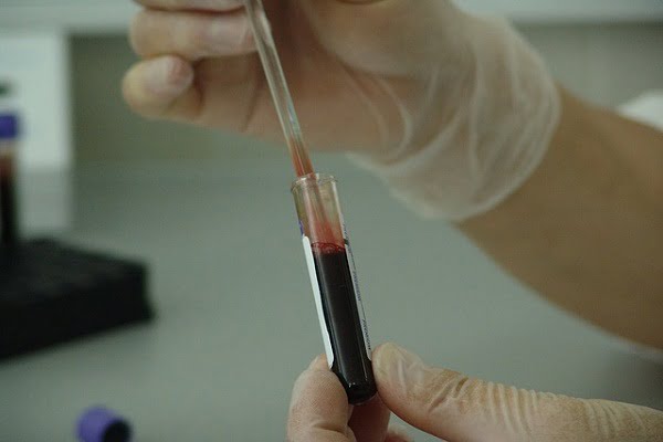
Your doctor can determine if you have abnormal levels of white or red blood cells or platelets. It is done by looking at a sample of your blood which may suggest leukemia. Various blood tests performed during diagnosis may include:
Complete blood count (CBC)
A CBC test is done to measure the number and quality of white blood cells, red blood cells and platelets as leukemia can cause abnormal blood cell counts.
Generally, immature blood cells called blasts or leukemia cells are not seen in the blood. Your doctor may suspect leukemia if there are blasts or blood cells that do not look normal in your blood.
Blood chemistry test
These tests are employed to measure level of certain chemicals in the blood. Blood level of these chemicals show how well certain organs are functioning and can help find the abnormalities. They are reliable sources of finding problems with the liver or kidney that may be caused by the spread of leukemia cells. Sometimes, these tests can also help the doctors to stage leukemia.
In leukemia, levels of the following chemicals may be higher than normal.
- Blood urea nitrogen (BUN)
- Uric acid
- Creatinine
- Phosphate
- Lactate dehydrogenase (LDH)
- Alanine aminotransferase (ALT)
- Aspartate aminotransferase (AST)
Bleeding and clotting factors
These tests measure the level of blood clotting factors in your blood to see how well your body can perform blood clotting. Usually, abnormal levels of blood clotting factors may occur with leukemia. Different tests used to measure these factors may include:
- Partial thromboplastin time (PTT)
- Prothrombin time (PT)
- Fibrinogen level
- International normalized ratio (INR)
Flow cytometry
It is a unique technique used to classify and sort the cells by labelling their surface with fluorescent labels. When these cells are exposed to a laser, it makes them give off a light. The light is then measured and analyzed by a computer.
Flow cytometry helps doctors to collect data rapidly from thousands of cells in a single sample and allows them to view many antibodies at the same time.
Flow cytometry can help to define and reveal the unique features of leukemia cells, or blasts. These features can then be used by the doctors to form a prognosis and measure response to treatment using minimal residual disease (MRD) accordingly. MRD usually refers to the presence of blasts in the bone marrow that can’t be found using standard lab tests such as microscopy, but are found using more sensitive tests such as flow cytometry or polymerase chain reaction.
Polymerase chain reaction (PCR)
Polymerase chain reaction (PCR) is an extra-sensitive test that is used to make many copies of a particular gene segment so that it can be tested in the lab. It helps measure the presence of certain biomarkers in blood or bone marrow cells.
The test measures minimal residual disease which refers to the remaining blood cancer cells which are not found by any other method generally. PCR can also be used to diagnose and check a patient’s molecular response to the treatment. It can specifically detect genetic abnormalities of the DNA or a specific marker found in patients with certain blood cancers such as acute chronic myeloid leukemia. It is also helpful in more sensitive follow-up of patients in remission of the cancer and can help to determine whether additional treatment is needed.
Bone marrow test for leukemia
Your doctor will perform a bone marrow test to examine the fluid and tissue in your marrow. This test is thought to be the confirmative test for leukemia as it helps in determining whether cancer or another disease is affecting blood cells or marrow, as well as the extent of the disease. Bone marrow test can help the doctor in early detection of changes in the blood cells before they can be seen in blood samples.
Bone marrow test usually involves two successive steps:
- bone marrow aspiration
- bone marrow biopsy
During a bone marrow aspiration, liquid part of marrow is removed and subjected to microscopic examination.
In a bone marrow biopsy, solid tissues of bone marrow are taken out and sent to a lab for further examination under a microscope by a pathologist. Both these tests are usually performed at the same time in a hospital establishment. The report from the lab confirms whether or not there are leukemia cells in the sample and, if so, which type of leukemia do they correspond to.
Lumbar puncture for leukemia
A lumbar puncture test is also known as spinal tap. In this test, a small amount of cerebrospinal fluid (CSF) that surrounds the brain and the spinal cord is removed from the space around the spine to look at it under a microscope.
To perform this test, your doctor will first numb an area in the lower part of the back over the spine. Then, he or she will place a small, hollow needle between the bones of spine and into the space around the spinal cord to collect some of the fluids. This test is performed to determine if the leukemia has spread into the spinal fluid.
Lymph node biopsy for leukemia
It is also known as excisional biopsy because it is a type of surgical biopsy where the whole of lymph node is removed. If the complete removal of lymph node is not possible, a portion of the lymph node is removed as a core biopsy.
After taking out either a portion or whole lymph node, it is sent to a lab for further examination under microscope by a pathologist to find out the presence of cancerous cells in the sample, identify the type of leukemia cancer and find out how quickly the cancer cells are growing.
Imaging tests for the diagnosis of leukemia
Imaging tests usually employ different forms of energy such as sound waves, x-rays, magnetic fields or radioactive particles and pass them through your body, creating pictures of the chest, abdomen, head, neck and other parts of the body.
Sometimes, your doctor may order an imaging test “with contrast,” which allows your doctor to see certain organs and tissues in the body more clearly. For this, before an imaging test is done, the technician will inject a contrast dye into one of your veins or a port or will ask you to drink a liquid containing the dye. If you’ve a history of reaction to the contrast dye or iodine in the past, you should tell your doctor or the technician before the examination.
Imaging tests are not helpful in the diagnosis of leukemia but they may be done for other reasons, such as to help in finding a suspicious area that might be cancerous. They may also be used to learn how far a cancer may have spread, or to help determine if the treatment has been effective.
Chest X-ray for leukemia
Small doses of radiations are used by an x-ray imaging system to capture an image of the body’s structures on a film. It is generally used to look for:
- lung infection (pneumonia)
- enlarged thymus gland
- enlarged lymph nodes in the centre of the chest (called mediastinal lymph nodes)
- buildup of fluid between the lungs and the walls of the chest (called pleural effusion)
Computed Tomography (CT) scan for leukemia diagnosis
A CT scan uses special x-ray device to create 3-D and cross-sectional images of organs, tissues, bones and blood vessels inside of your body. A computer then recombines these images and turns into a detailed set of pictures.
A CT scan may be used to look for enlarged spleen and liver. It is also helpful in looking and determining if the lymph nodes around the heart, near the trachea (windpipe) or in the back of the abdomen are enlarged.
More: What is an enlarged heart?
Magnetic resonance imaging (MRI) scan for diagnosing leukemia
MRI uses radiofrequency waves and powerful magnetic forces to create cross-sectional images of organs, tissues, bones and blood vessels of your body. A computer then recombines these images and turns them into 3-D pictures. It takes longer time to perform an MRI scan than a CT scan. It may require up to an hour.
MRI is most useful in looking brain and spinal cord and it is often used when doctors think that the leukemia has spread into the brain.
Ultrasound for leukemia
Ultrasound uses high-frequency sound waves to create images which can be used to look at lymph nodes near the surface of the body or to look for enlarged organs (like the liver and spleen) inside your abdomen and whether these organs have been affected by the leukemia.
This is relatively an easy test to do. It doesn’t use any kind of radiation. During this procedure, you will be required to simply lie on a table, and a technician moves the transducer over the part of your body being looked at.
Treatment for leukemia
Your doctor will prepare a customized treatment plan for you after the diagnosis of leukemia is established. Treatment plan is based on your specific needs and it may include a combination of different treatment options. While deciding about the treatment plan, your doctor may consider these factors.
- The type of your leukemia
- Your age
- Chromosomal (genetic) abnormalities
- Your overall health
Chemotherapy for leukemia
Chemotherapy is thought to be the standard treatment for many types of leukemia. If the cure of leukemia is not possible, still it can help you live longer and feel better so as to improve the quality of life.
Usually, a combination of anti cancer drugs is used in chemotherapy for leukemia. Combinational administration of drugs attack leukemia cells in different ways and it also helps to stop the leukemia cells from becoming resistant to any one drug.
For better working of chemotherapy drugs and to prevent infection or bleeding during the treatment, various other medicines may be given along with chemotherapy drugs. Example of these drugs include epoetin and hematopoietic stimulants.
Sometimes, many types of acute leukemia may spread to your brain and spinal cord. Regular chemotherapy is unable to reach those areas because your body puts up a special barrier to protect them. For these kinds of leukemia, intrathecal chemotherapy is applied which treats these areas by attacking any leukemia cells present there. In this treatment, anti-cancer drugs are injected directly into your spinal canal.
Chemotherapy drugs used in different kinds of leukemia are listed below.
For acute lymphoblastic leukemia (ALL), your doctor may recommend one or a combination of the following drugs.
- Asparaginase
- Blinatumomab
- Clofarabine
- Daunorubicin
- Doxorubicin
- Methotrexate
- Nelarabine
- Vincristine
- Corticosteroids such as dexamethasone or prednisone
Acute myelogenous leukemia (AML) may be treated with chemotherapy medicines which may include:
- Cytarabine
- Daunorubicin
- Idarubicin
- Mitoxantrone
For chronic lymphocytic leukemia (CLL), the following drugs may be used as chemotherapy medicines.
- Bendamustine
- Chlorambucil
- Cyclophosphamide
- Fludarabine
- Vincristine
- Corticosteroids such as prednisone
- Monoclonal antibodies such as alemtuzumab or rituximab
For chronic myelogenous leukemia (CML), these drugs may be used as chemotherapy medicines by your doctor.
- Cyclophosphamide
- Cytarabine)
- Tyrosine kinase inhibitors such as dasatinib, imatinib, or nilotinib
Patients having CML who can’t take tyrosine kinase inhibitors and are unable to receive stem cell transplants, may be administered busulfan, hydroxyurea, or interferon alfa with or without cytarabine.
Chemotherapy is generally associated with lots of side effects which primarily depend on the type and dose of the drug being taken or the regimen of the drug. Some of the common side effects of chemotherapy may include:
- Hair loss
- Nausea
- Vomiting
- Mouth sores
- Loss of appetite
- Tiredness
- Easy bruising or bleeding
- Increased chance of infection due to the destruction of white blood cells
Your doctor may prescribe medicines to mitigate these side effects of the chemotherapy. It has been observed that some adult men and women who receive chemotherapy sustain damage to the ovaries or testes, resulting in infertility. Though, most of the children who receive chemotherapy for leukemia will have normal fertility as adults but depending on the kind and dose of the drug being used, some may experience infertility as adults.
Biological therapy for leukemia
Biological therapy refers to the use of artificial or natural substances that change the way cells behave. Different agents of biological therapy work in different ways. Some agents are used to kill, control or change how cancer cells behave while some other agents are responsible for strengthening the body’s immune system, control symptoms or lesson the side effects of treatment. Biological therapy is known by some other names such as biotherapy or biological response modifiers (BRMs).
Biological therapy is mainly used to treat chronic myelogenous leukemia (CML) where prime objective of biological therapy is:
- to return blood cell counts to normal when CML is in chronic or accelerated phase
- to treat CML relapses after a stem cell transplant
Biological therapy may have its side effects but they tend to be less severe than those of chemotherapy and can include rash or swelling at the injection site for IV infusions of the therapeutic agents.
Headache, muscle aches, fever, and tiredness are some other side effects experienced by patients having biological therapy.
Targeted therapy for leukemia
Unlike standard chemotherapy where all cells in the body are affected, targeted therapy uses drugs that attack specific vulnerabilities within leukemia cells, helping to reduce the damage to healthy cells and minimize the side effects. Targeted therapy may be used as a standalone treatment option or in combination with other leukemia treatments, such as chemotherapy.
For example, the drug imatinib is used in people with chronic myelogenous leukemia (CML) which stops the action of a protein within the leukemia cells of CML. This helps in controlling the progression of the disease.
Although targeted therapy has less side effects than chemotherapy but it may cause side effects such as reduction in blood counts, which may lead to fatigue and risk of infection. Other general side effects of targeted therapy may include fever, chills, headache, rash, nausea and shortness of breath.
Generally, targeted therapies are given either in a pill form or through injection into your veins. Some other common side effects of targeted therapy may include swelling, bloating, sudden weight gain, nausea, vomiting, diarrhoea, muscle cramps and skin rash.
Radiation therapy for leukemia
Radiation therapy uses X-rays or other high-energy beams. This is mainly used to prevent leukemia from spreading to, or treat leukemia that has spread to, the central nervous system (CNS) by damaging leukemia cells and stop their growth.
During a radiation therapy, you simply lie on a table and a large machine moves around you, directing the radiation beams to precise points on your body where a collection of leukemia cells is confirmed through diagnosis. You may also receive the radiation therapy over your whole body. Radiation therapy is also used to prepare your body for a bone marrow transplant.
Radiation therapy either destroys the cancer cells or shrinks the tumours. Sometimes, in leukemia, it is used to shrink the swollen lymph nodes.
Radiation therapy may cause temporary side effects which go away once the treatment is stopped. Usually, the side effects of radiation therapy depend on the location of the body that is irradiated. For example, you may experience symptoms like nausea, vomiting, and diarrhoea if the radiation is given to your abdomen.
One of the most common side effects with any kind of radiation therapy is related to the skin in the area being treated. The skin there may become red, dry, and tender. While undergoing radiation therapy, you may feel a generalized tiredness persistently.
Stem cell transplant for leukemia
A stem cell transplant, sometimes referred as bone marrow transplant, is a procedure to replace your unhealthy bone marrow with healthy bone marrow which results in replacement of unhealthy blood-forming cells with healthy cells. It allows your body to receive large doses of chemotherapy or radiation therapy to increase the chance of eliminating blood cancer in the marrow and then restoring normal blood cell production. However, continuous researches are going on to improve stem cell transplantation procedures, making them an option for more patients.
Before you will go for a stem cell transplant, your doctor will give you a high dose of chemotherapy or radiation therapy to destroy your diseased bone marrow including the leukemia cells. Then the doctor will infuse blood-forming stem cells to your body that help to rebuild your bone marrow.
The basis for stem cell transplantation is that stem cells give rise to different blood cells such as red cells, white cells and platelets, which are present in the marrow, peripheral blood and cord blood. High dose chemotherapy or radiation therapy kills the patient’s stem cells which stops the stem cells from making enough blood and immune cells.
If you are a potential candidate for a stem cell transplant, your doctor may recommend one of the following types of stem cell transplant.
- Autologus – the new stem cells used in this type of transplant are from your own body
- Allogenic – the new stem cells are taken from a healthy person usually known as donor
- Reduced intensity stem cell transplantation – like allogenic transplant, the stem cells are taken from a healthy donor but the chemotherapy given in this case is less intensive
- Syngeneic transplantation – it is a rare type of stem cell transplant which is only used on identical twins where donor is one of the identical twins
Complications with stem cell transplant for leukemia
Complications related to stem cell transplant include infections and bleeding due to the depletion of normal blood cells.
A major risk of stem cell transplant with donor cells is known as graft-versus-host disease (GVHD) which can be both mild and severe in its intensity. In GVHD, a patient’s normal tissues are attacked by the white blood cells of the donor. It generally affects your liver, skin, or digestive tract. GVHD may precipitate itself at any time after the transplant, even years later. Medications that suppress the immune response or steroids are generally used to treat this condition.
Outlook and Prognosis: What is the prognosis and survival rate with leukemia?
The prognosis of leukemia largely depends upon the type of leukemia that is present, health status of the patient and the age of the patient. Death


