What is uterine or womb cancer?
Uterine or womb cancer is one of the most common cancers of the female reproductive system. It is a malignant tumor that occurs in the cells of the uterus and also can spread or metastasize to other parts of the body.
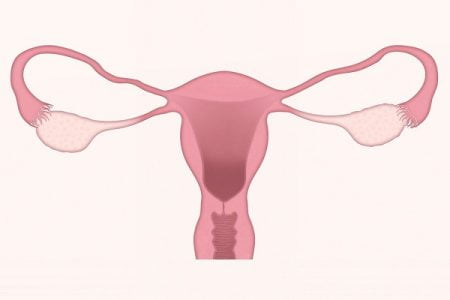
The uterus or womb is an important part of a woman’s reproductive system where a fetus develops and grows during pregnancy. The uterus is a hollow, muscular and pear-shaped organ. The uterus is covered by a lining called as endometrium which is made up of many tissues and glands.
The lower most part of the uterus is called cervix which leads into the vagina. Sometimes the cells in the uterus undergo some changes and thereafter do not grow or behave normally. These changes may lead to non-cancerous and benign conditions like endometriosis or non-cancerous tumors like uterine fibroids.
The changes in uterine cells can also result in pre-cancerous conditions where the abnormal cells are not yet cancer but there are chances that these may become cancer if not treated. For example, one of the most common pre-cancerous conditions of the uterus is atypical endometrial hyperplasia.
The changes in the uterine cells can sometimes result in the formation of cancerous tumor in the uterus.
Types of uterine cancer
There are three main types of uterine cancer:
- Endometrial cancer
- Uterine sarcoma
- Choriocarcinoma
Endometrial cancer
Cancer of the uterus which develops in the lining of the uterus (endometrium) is called endometrial cancer. Almost all uterine cancers start in the endometrium. There are two main types of endometrial cancer:
- Endometrioid adenocarcinoma
- Uterine carcinosarcoma
Endometrioid adenocarcinoma
Endometrioid cancer is the most common type of adenocarcinoma and accounts for most cases of endometrial cancers. Endometrioid adenocarcinoma cancers are made up of cells in glands that resemble the normal uterine lining (endometrium). Some of these cancers consist of both squamous as well as glandular cells.
Uterine carcinosarcoma
Uterine carcinosarcomas consist of cancer cells which resemble endometrial cancer and sarcoma. These endometrial cancers have a high risk of spreading to the lymph nodes and other parts of the body.
Uterine sarcomas
Uterine sarcomas are common types of uterine cancer which start either in the muscle wall of the uterus (myometruim) or in the connective tissue supporting the endometrium (stroma). Uterine sarcomas are usually rare tumors comprising almost 4.5% of all malignant growths of the uterus and nearly about 1–3% of genital tract cancers. These tumors develop most frequently in women between the ages of 40 and 50 and are rare before the age of 30. Uterine sarcomas are of four main types:
- Intramural uterine sarcomas
- Mucosal uterine sarcoma
- Leiomyosarcoma
- Endometrial stromal sarcoma
Intramural uterine sarcomas
In intramural sarcomas, the tumors arise in the myometrium layer of uterus.
Mucosal uterine sarcoma
Mucosal uterine sarcoma tumors develop from the endometrium of the uterus.
Leiomyosarcoma
Uterine leiomyosarcoma develops in the myometrium layer of the uterus and is the most common type of uterine sarcoma.
Endometrial stromal sarcoma
This is a rare type of uterine sarcoma which develops in the supporting connective tissue of the endometrium. Low-grade stromal sarcomas are mostly found in pre- menopausal women while high-grade stromal sarcomas are found in postmenopausal women.
Choriocarcinoma
Choriocarcinoma is a rare type of uterine cancer, however it is one of the most malignant growths that develop in the body of the uterus. This rare type of tumor mostly affects pregnant women and is called gestational choriocarcinoma. This type of uterine cancer usually starts in the uterus but can also spread to other parts of the body. Choriocarcinoma is actually a type of gestational trophoblastic disease (group of rare tumors which involve abnormal growth of cells inside the uterus.
Signs and symptoms of uterine cancer
The main signs and symptoms of uterine cancer include:
- Abnormal discharge or bleeding from vagina
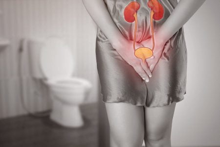
- Difficulty in urination
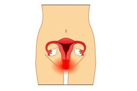
- Pain in the pelvis
- Painful intercourse
- Pain in the lower part of abdomen
- Pain in the back and legs
- Appetite loss
- Excessive tiredness
- Nausea
- Unusual weight loss
- Unusual heavier flow during periods
- Vaginal bleeding even after menopause
- Abnormal and watery blood-tinged discharge from the vagina
- Breathing problems
- Difficulty in bowel movement
- Presence of blood in the stools
- Accumulation of fluid in the abdomen (ascites) and in the legs (lymphedema)
- Excessive bleeding from the rectum or bladder
- Feeling of pressure in the pelvis
- Feeling of fullness in the abdomen or pelvis
- Presence of a mass (tumor) in the uterus which can be easily felt
- Recurrent urination
- Nausea
- Bloating
- Changes in bowel and bladder habits
- Anemia due to excessive blood loss
- Cramping in the abdomen or pelvis
Causes of womb/uterus cancer
The exact cause of uterus cancer is unknown yet, however following factors play an important role in the development of uterus or womb cancer:
Age
Uterus cancer risk usually increases with age and in most of the cases it occurs in women aged between 40 to 74 years and almost 1% of cases have been diagnosed in women under the age of 40.
Oestrogen levels after the menopause
The uterus cancer has been linked to the body’s exposure to oestrogen which is one of the hormones that help in the regulation of reproductive system in women. The levels of oestrogen in the body are usually maintained by hormone progesterone. If oestrogen levels are not maintained by progesterone, its levels can increase, called unopposed oestrogen.
After menopause, progesterone is not produced by the body, however, small amounts of oestrogen are stll produced. As a result this unopposed oestrogen divides the cells of the endometrium and therefore increases the risk of womb cancer.
Obesity
Oestrogen can be also produced by fat tissues in the body, so being overweight or obese increases the levels of oestrogen in the body which in turn increases the chances of developing womb cancer. It has been found that women who are overweight are 3 times more likely to develop womb cancer as compared with women who have a normal healthy weight. Very obese women are 6 times more prone to develop womb cancer when compared with women of normal healthy weight.
Reproductive history
It has been found that women who have not had children are at a higher risk of developing womb cancer.
Tamoxifen
It has been found that women taking tamoxifen (a hormone used for the treatment of breast cancer) are at an increased risk of developing womb cancer.
High insulin levels
Conditions such as hyperinsulinemia, in which body produces more insulin than normal, increase the risk of womb cancer.
Polycystic ovary syndrome (PCOS)
Women with polycystic ovary syndrome (PCOS) have high levels of oestrogen in their bodies, so they are at a higher risk of womb cancer.
Endometrial hyperplasia
Endometrial hyperplasia, abnormal thickening of endometrium (inner lining of the womb) also increases the risk of womb cancer.
Uterine cancer risk factors
Risk factors of uterine cancer include the following:
- Obesity
- High fat diet
- Age
- Tamoxifen
- Personal or family history of uterine, ovarian or colon cancer
- Diseases in the ovaries like polycystic ovarian syndrome (PCOS)
- Atypical endometrial hyperplasia
- Diabetes
- Early menstruation (before 12) and late menopause
- Breast cancer
- Ovarian cancer
- High dose radiation therapy in the pelvic area during the treatment of other types of cancer
Diagnosis: How is uterine cancer diagnosed?
If any woman has the symptoms of uterine cancer described above, she should immediately consult a doctor for proper diagnosis.
Following are some of the procedures that might be used in the diagnosis of uterine cancer in women:
Endometrial biopsy
In this procedure, a sample of endometrial tissue is obtained with the help of a very thin flexible tube that is inserted into the uterus through the cervix. The tube removes a small amount of the endometrium sample using suction.
Transvaginal ultrasound or sonography
A transvaginal sonogram is used to create pictures of the uterus with the help of sound waves.
Dilation and curettage (D & C)
In this procedure, a special surgical instrument is used to scratch tissue from inside the uterus and increasingly. This procedure is done in a special operating room.
Testing of endometrial tissue
Endometrial tissue samples that are removed by biopsy or D & C are analyzed under the microscope to find out whether cancer is present or not.
CT or CAT scan (computerized tomography or computerized axial tomography)
This scan involves taking a series of pictures of areas inside the body with the help of a computer linked to an X-ray machine. These images are then used to check any abnormality of tumor present in the body. It can also help to determine the size of the tumor.
MRI (magnetic resonance imaging)
In this procedure, radio waves and a powerful magnet linked to a computer are used to create pictures of areas inside the body. MRI helps in measuring the size of tumor. MRI is often useful in women with low-grade cancer to check how far the cancer has developed in the wall of uterus.
Positron emission tomography (PET)
This test helps in spotting collections of cancer cells. It consists of special scanners which can be combined with CT to locate the areas of cancer spread more precisely. PET scans are not routine diagnostic tests in detecting uterine cancers; however they are used in more advanced cases.
Blood tests
Some of the main blood tests used in the diagnosis of uterine cancers are:
Complete blood count (CBC)
The CBC test is used to measure different types of cells present in the blood like RBCs, WBCs and platelets. Due to excessive bleeding in uterine cancers, the RBC count falls low (anemia) and can be analyzed by CBC.
CA-125 assay
This blood test is used to measure the level of CA-125, a substance that is occasionally found in an increased amount in the blood, other body fluids, or tissues. A particular level of CA-125 recommends the presence of some types of cancer.
Treatment options for uterine cancer
Uterine cancer is usually treated by a combination of treatments like surgery, radiation therapy, chemotherapy, and hormone therapy. Combination of treatments depends on the stage and distinctiveness of the cancer. Treatment options usually depend on various factors like type and stage of cancer, overall health of patent, age and possible side effects. Following are some of the treatment options:
Surgery for uterine cancer
Surgery is the first treatment used for uterine cancer and involves the removal of the tumor and surrounding healthy tissue, called as margin, during an operation. Commonly used surgical procedures for uterine cancer are:
Hysterectomy
Depending on the level of the cancer, the surgeon will perform either a simple hysterectomy (removal of the uterus and cervix) or a radical hysterectomy (removal of the uterus, cervix, upper part of the vagina and some nearby tissues). A typical bilateral salpingo-oophorectomy is usually recommended which is the removal of both fallopian tubes and ovaries.
Lymphadenectomy
It is a procedure of removing lymph nodes near the tumor site in order to determine if the cancer has spread beyond the uterus or not.
Radiation therapy
Radiation therapy uses high-energy x-rays and other particles in order to destroy cancer cells. Radiation therapy along with surgery is given to some women with uterine cancer. Radiation therapy is sometimes given to patients before surgery to shrink the tumor.
Chemotherapy
Chemotherapy is usually given after surgery along with radiation therapy and involves use of drugs to destroy cancer cells. It usually acts by ending the ability of cancer cells to grow and divide. Chemotherapy is also given if the uterine cancer returns after initial treatment.
Hormone therapy
Hormone therapy slows down the growth of various types of uterine cancer cells. These tumors are usually adenocarcinomas and belong to either grade 1 or grade 2 tumors. Hormone therapy in uterine cancer usually involves a high dose of the sex hormone progesterone given in tablet form. Hormone therapy is usually given to women who cannot undergo a surgery or radiation therapy. It can be also used in combination with other types of treatment.


