Brain Cancer (Brain Tumor): Causes, Signs, Symptoms, Diagnosis, Prevention, Treatment, Stages, Survival Rate
- Updated on: Jul 10, 2024
- 20 min Read
- Published on Sep 26, 2019


What is brain cancer (brain tumor)?
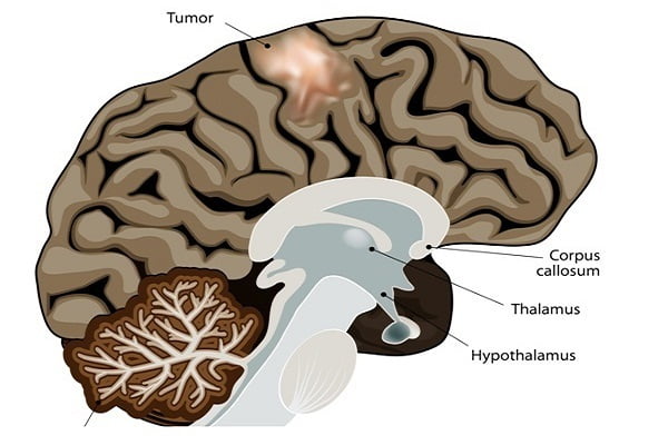
Brain cancer is the formation of tumours in the brain that have grown out of control. Although many growths in the brain are generally called brain tumors, but not all brain tumors are cancerous.
Tumours formed in the brain can be of two types – Benign tumours, also called non-cancerous tumours and malignant tumours which are cancerous in nature. The main difference between benign and malignant tumours is that benign tumours do not grow into the nearby tissues (or spread to distant areas of your body) while malignant tumour does. Benign tumours are generally not life-threatening while malignant tumours are dangerous.
It is rarely observed that brain tumours spread to other parts of the body; however most of them can spread through the brain tissue. In some cases, as the benign tumours grow in size, they press on and destroy normal brain tissue causing damage that is often disabling and sometimes may be fatal.
The term, brain cancer, is specifically reserved for malignant tumours of the brain. Malignant tumours are composed of aggressively growing and abnormal-appearing cells referred to as cancer cells. The main aspect of brain tumours that doctors usually focus on is how readily they can spread through the rest of the brain and whether they can be removed with little or no chances of their coming back.
Malignant tumours can disrupt the way your body works, which can be life-threatening. However, brain cancer is quite uncommon. American Cancer Society estimates that your chance of developing a malignant brain tumour in your lifetime is less than 1 percent.
Brain tumours tend to be of different nature in adults and children. They often develop from different cell types, form in different areas and may have different outlook and require different treatment.
Primary and secondary brain tumours
Brain tumours are classified on the basis of their origin. If they are arising from primary brain cells, the cells that form other brain components (for example, membranes, and blood vessels), the tumor is known as primary brain tumour. Primary tumours are further classified and named after the part of the brain or the type of brain cell from which they arise.
If the tumour in brain has formed due to the spread of cancer cells that develop in other organs, the tumor is known as metastatic or secondary brain tumour. Metastatic brain tumours are the most common types of the brain tumours.
Brain aneurysm Vs Brain tumours
Sometimes brain tumours are confused with brain aneurysm. Brain aneurysms are expanded areas in the brain arteries or veins that are abnormally weak and form a ballooning or expansion of the vessel wall. Generally, they do not produce any symptoms unless they begin to leak blood into the surrounding brain tissue or they burst. Brain aneurysm is not a tumour.
Aneurysms may be present at birth – congenital or have formed in brain vessels after vessel damage in case of trauma, atherosclerosis or high blood pressure. They are never formed from cancer cells. However, when aneurysms produce symptoms, they can resemble the symptoms manifested by the brain tumours.
Paediatric brain tumours
Brain tumours in children are different than tumors in adults due to the fact that their bodies and brains are still developing. The prevalence of the most common childhood brain tumours is different from the most common brain tumours found in adults.
Though brain cancer is rare as such but it is the most common form of solid tumours among children under the age of 15 and also among one of the leading causes of deaths due to cancer.
Paediatric brain tumours can occur in different locations and behave differently than brain tumours in adults. Treatment strategy for brain tumours in children largely depend on the type of tumour and age of the child. Generally, children have much better prognosis than adults with a similar condition of tumour.
How common is brain cancer?
Presently, an estimated 700,000 Americans are living with brain tumour, of which 80% tumours are benign and the remaining tumors are malignant.
In 2018, it is estimated that about 79,000 people will be diagnosed with primary brain tumour out of which more than 55,000 will be benign tumours and about 24,000 will be malignant tumours. It is also estimated that about 17,000 people will die from brain cancer in 2018.
The average survival rate for all malignant brain tumour patients is only 34.7%. For males, it is found to be 33.8% and for females it is 36.4%.
What are the causes of brain tumour (brain cancer)?
Like any other cancer, the exact cause of brain tumour development is unknown. Also, the well established risk factors for brain tumours are very few in number. Researchers suggest that DNA changes or mutation in normal brain cell might be a possible reason that leads to the development of brain tumours. The brain cells divide rapidly after mutation and form a mass known as tumour after the uncontrolled cell division.
These gene changes can be of two types. Inherited gene changes are passed from parents to the offspring while the other types of changes are acquired during a person’s lifetime due to various factors such as tobacco use and cigarette smoking etc.
Brain tumor risk factors – What are the chances of getting a brain tumor?
Risk factors affect your chances of getting brain tumours. There are some risk factors which can be controlled such as use of tobacco, and there are some others which are simply out of your control such as genetic changes or family history that can’t be changed. However, you should know that the mere presence of one or more of these risk factors does not mean you will develop the brain tumour. Many people get this disease without having any known risk factor. Similarly, not having any risk factors does not mean you will never get the tumor.
There are some risk factors which make you more susceptible to developing brain tumours. These may include the following.
Age
A brain tumour can occur at any age; however brain tumours are more common in age between 60 and 80 years. There are some types of brain tumours which are more common in children and young adults.
Gender
Usually, men are more likely to develop a brain tumour than women. However, there are some specific types of brain tumours which are more common in women such as meningioma.
Exposure to various types of radiation
Radiation exposure is the prime known environmental risk factor for brain tumours. It is most commonly encountered in radiation therapy during treatment of some other disease or medical conditions. For example, earlier, children with a fungal infection of the scalp were sometimes treated with low dose radiation therapy. This was later found to have increased the risk of brain tumours on ageing.
Most of the radiation-induced brain tumours occur due to the treatment of other cancers around head using radiations. Children who get radiation as part of their treatment for leukaemia are the most vulnerable population to develop radiation-induced brain tumour. It usually develops around 10 to 15 years after the radiation treated is delivered.
Risk of radiation exposure due to imaging tests such as x-rays or CT scans have no confirmed effect on the development of brain tumour. Since the radiation level used in these tests are much lower than those used in radiation therapy, the risk for brain tumour, if any, is thought to be very small. For safety concerns, doctors do not recommend even these tests unless it is very necessary.
Family history
Brain tumours due to family history are very rare, but having family history of certain diseases can almost double the risk of brain tumours. Some of the familial diseases which increase the risk of brain tumors or cancers are:
- Neurofibromatosis type 1 (NF1)
- Neurofibromatosis type 2 (NF2)
- Tuberous sclerosis
- Von Hippel-Lindau disease
- Li-Fraumeni syndrome
Other brain tumor risk factors
Cancer of other parts of the body can also spread to the brain. Cancer that usually metastasize to the brain and causes development of secondary brain tumours include breast cancer, lung cancer ,heart cancer ,bladder cancer and melanoma or the cancer of the skin.
Exposure to certain chemicals, solvents, pesticides, oil products, rubber or vinyl chloride at home or work may also increase the risk of brain cancers but no definite evidence is available in the scientific community for this claim.
Serious head injury or trauma has also been linked to the development of brain tumours, particularly the meningoma type of brain tumours, in some studies.
What are the various signs and symptoms of brain cancer?
Not all brain tumours manifest their early symptoms, and many times, the tumour is found only after the death of a person. The death may have happened due to other illness or disease also and not necessarily due to brain cancer. Sometimes, people do not recognize that they have brain tumour, because the general symptoms may resemble other medical conditions or diseases. However, if brain tumor symptoms are visible, they can be of two types – general or specific.
General brain tumor symptoms are caused due to the pressure or encroachment of tumor on other parts of the brain or nearby structures. The specific symptoms are caused by the location of the brain tumour itself, which affects the normal functioning of that specific part of the brain.
What are the general symptoms of brain cancer?
General symptoms of the brain cancer include:
- Headaches – often get worse in the morning, may become persistent and severe
- Nausea and vomiting
- Muscle weakness
- Reduced touch sensations
- Problem with coordination and balance, clumsiness
- Fatigue of arms and legs
- Difficulty in walking
- Seizures
- Difficulty with speech or impaired voice
- Vision abnormalities such as blurred vision, double vision and loss of peripheral vision
- Personality changes
- Change in intellectual and emotional capacity
- Memory problems
- Change in concentration and alertness
- Decline in brain function or confusion
- Muscle jerking and muscle twitching
It should be remembered that many of these symptoms appear gradually and can be overlooked for a long time before they are detected. You should consult your doctor if you are having these symptoms persistently to diagnose the cause and treat it accordingly.
What are the various types of brain cancers or brain tumours?
The name of a cancer type is largely derived from the location of your body where the cancer begins. The cancer beginning in various brain parts or its related structures is known as brain cancer. Brain cancer can begin in any part of your brain and is named accordingly.
The type of cancer beginning from your brain is referred as primary brain cancer. Some cancers start elsewhere in your body and then reach to the brain through metastasis and are known as secondary brain cancer or metastatic brain cancer. Large number of tumours found in brain is because of metastatic tumours rather than primary brain tumours.
Primary brain tumours can be further classified based on the invasion potential to affect nearby tissues – least invasive being called grade I brain cancer and the most invasive being called grade IV brain cancer. Brain tumors can also be classified on the basis of their nature such as benign primary tumours which are non cancerous, and cancerous brain tumours which are cancerous in nature. The grades of brain tumours are assigned based on the type and location of the tumour. Grades also refer to the growth rate of the tumour or how quickly the tumor or cancer can grow.
There more than 120 types of reported brain tumours. Many of them have further sub-types. Lack of a decisive classification infrastructure results in different doctors using different names for the same tumour.
Some of the most common primary brain tumours are discussed below.
Gliomas
Any cancer that starts from the glial cells of the brain is known as gliomas. A glial cell functions as supportive cell in your brain. Glioma is a general term used to refer to the cancer arising from these cells rather than of any specific type of cell. Various subtypes of gliomas include:
- Astrocytomas (which include glioblastomas)
- Oligodendrogliomas
- Ependymomas
About 30% of primary brain tumours are gliomas. Gliomas are the most fast-growing brain tumours as found in researches.
Astrocytomas
Astrocytomas are tumours that start in the cells called astrocytes. Astrocytes are the type of glial cells found in your central nervous system. Astrocytomas are the most common type of gliomas. It is very rare for them to spread outside of the brain but sometimes they do spread to the cerebrospinal fluid (CSF) pathways. There are four grades of these tumours based on how quick they grow and how aggressive they are.
- Grade I astrocytoma is a slow growing and benign brain tumour which rarely spreads to the nearby tissues. This is more commonly found in children. It is also known as pilocytic astrocytoma.
- Grade II astrocytoma is a low-grade brain tumour which often spreads to nearby tissues. These are slow growing tumours. These tumors are capable of transforming into higher grade astrocytomas.
- Grade III astrocytoma is called anaplastic astrocytoma. This type of tumour grows quickly and spreads to the nearby tissues. It is a cancerous brain tumour.
- Grade IV astrocytoma is also known as glioblastoma which is the most advanced and aggressive form of astrocytoma.
Oligodendrogliomas
These tumours start in oligodendrocytes which is a type of glial cells of brain. These tumours tend to grow slowly and most of them can invade the nearby brain tissues. These are very hard to remove completely by surgery.
Sometimes these tumours spread along the CSF pathways but rarely spread outside the brain. With time they become very aggressive and transform into high grade tumours. Oligodendrogliomas constitutes only about 2% of brain tumours.
Ependymomas
These tumours start in the lining cells of the ventricles of your brain known as ependymal cells. They are usually found to be in range of grade II to grade III. These tumours are more likely to spread to the cerebrospinal fluid pathways and block the exit of CSF from the ventricles of your brain. This blocking results in ventricles enlargement – a medical condition known as hydrocephalus.
They very rarely grow into normal brain tissue. These types of tumours can be cured and removed completely with the help of surgery but there are chances of nerve damage during the surgical process to remove it.
Meningiomas
This kind of tumour arises from the meninges. Meninges are the membranous layer of tissues which surrounds around the outer part of your brain and spinal cord. It is the most common type of brain tumour in adults and accounts for every 1 out of 3 primary brain tumours.
Most oftenm meningiomas do not cause any symptom even after growing for many years because of their slow growth rate. But sometimes, they result in serious disability of adjacent brain tissues, nerves and blood vessels.
Women are more prone to this kind of brain tumours. It is more common in old-aged people, though it can occur at any age.
Medulloblastomas
Medulloblastomas are the cancerous brain tumours (malignant) that develop from neuroectodermal cells (early forms of nerve cells) in the lower back part of your brain called the cerebellum. Brain tumours of this category are fast growing and often spread throughout the CSF pathways.
Medulloblastomas are more common in children than in adults. They belong to a class of tumours called embryonic tumours that that start in the embryonic cells in the brain.
Medulloblastoma can be treated with the help of surgery followed by radiation or chemotherapy or both as adjuvant therapy.
Gangliogliomas
Gangliogliomas are the brain tumours found in both the neuronal and glial cells of the brain. It is a rare kind of brain tumour and is very uncommon in adults. When found, they are characterized by their typical slow growth rate. They are usually cured by surgery alone or surgery combined with radiation therapy.
Craniopharyngiomas
Craniopharyngioma is a rare type of benign (non-cancerous) brain tumour that starts just above the pituitary gland. It is able to press over the pituitary gland and hypothalamus which affects the normal function of these structures resulting in hormonal problems in your body. These tumours can also be cause vision problems because they occur in the proximity of optic nerves in your brain.
Majority of the cases of these tumours are found in children and older adults, though they can occur at any age. Children with this type of brain tumour are found to encounter slow growth rate and smaller than average body’s overall growth.
Schwannomas
Schwannoma is a rare type of brain tumour that begins in the Schwann cells of the nerve sheath, which surrounds and insulates the cranial nerves and other nerves. Schwannomas constitute about 8% of all tumours found in the central nervous system which includes your brain and spinal cord.
Mostly, these tumours are benign or noncancerous in nature and are found to occur in the vestibular nerve. Vestibular nerve is present in the inner ear which helps in controlling and coordinating your body balance. Schwannomas of vestibular nerve is also known as acoustic neuromas.
Pituitary tumours
The tumours which start in your pituitary gland as abnormal growth are known as pituitary tumours. These tumours are benign or non-cancerous known as adenomas. These tumours sometimes result in overproduction of hormones by your pituitary gland and can, in some cases, even decrease the normal level of hormone production which regulates important functions of your body. Adenomas of pituitary gland lack the malignancy power and remain localised in the gland and surrounding tissues. They typically do not spread to other parts of the body.
Pituitary tumours are treated with various different approaches which may include removing the tumour, controlling its growth and managing your hormone levels with medications.
Grades of brain tumours – Brain cancer Grading
Assigning a grade to the brain tumour signifies its seriousness and extent to which it has advanced. Tumour grade is assigned based on the tumour’s microscopic appearance in comparison to normal brain cells using some or all of the following criteria:
- similarity to normal cells
- growth rate or mitotic index
- indications of uncontrolled growth
- tumour necrosis or presence of dead cells in the centre of the tumour
- possibility to spread or potential for invasion based on whether or not it has a definitive margin (diffuse or focal)
- blood supply to the tumour or vascular environment of the tumour
Most doctors use the World Health Organization (WHO) classification system to grade brain tumours which is explained below.
WHO Grading System for brain tumours
Grade I Tumour
- have slow growth rate of tumour cells
- appear almost normal under a microscope
- are least malignant
- survival is usually long-term ?
Grade II Tumour
- Relatively slow-growth of tumour cells
- Appear slightly abnormal under a microscope
- Have potential to invade adjacent normal tissue
- Can transform into higher grade tumour
Grade III Tumour
- Actively reproducing abnormal cells
- Appears very abnormal under a microscope
- Infiltrate adjacent normal brain tissue
- Tumour tends to recur, often as a higher grade
Grade IV Tumour
- Abnormal cells which reproduce rapidly
- Very abnormal appearance under a microscope
- Form new blood vessels to maintain rapid growth
- Necrotic cells are present in the centre of the tumour
Diagnosis of brain cancer
If your doctor suspects a brain tumour after manifestation of any of the symptoms, he/she will take your thorough medical history and will conduct a physical examination. The doctor will also perform your neurological examination to check your brain function. In a neurological examination, your doctor may test your reflexes, vision, eye and mouth movement, muscle strength, balance, coordination, alertness and other such functions.
During physical examination, your doctor might gently test your breasts, tummy and skin for any visible signs of cancer of these places.
If the results of a physical examination are abnormal, your doctor will recommend you to visit a neurologist who will perform different more tests to establish a confirmed diagnosis of the brain tumour.
These tests may include:
- Imaging tests
- Brain biopsy
- Lumbar puncture
- Blood and urine tests
Imaging tests for brain tumour
Different imaging test produce three dimensional image of your inside of your body using principles of x-rays, sound waves and magnetic waves. These images can be used to look at a tumour, its location and size. Imaging test can also help in treatment plan for different types of tumours.
The two most used and useful test for your brain tumour are:
- Magnetic Resonance Imaging (MRI) Scan
- Computed topography (CT) Scan
Magnetic Resonance Imaging (MRI) Scan
MRI scans are considered the best way to look for tumours in the brain as it is very good looking at it. The images they provide are usually more detailed than those from CT scans (described below). But they do not image the bones of the skull and therefore may not show the effects of tumours on the skull.
MRI scanning is a commonly used technique to diagnose brain tumours. It uses radio waves and strong magnets to generate pictures to details of the brain. The image obtained by this technique is usually more detailed than other imaging test. One of the disadvantages of this test is that it cannot image the bones of the skull.
There are various types of MRI scans to image specific locations of the brain and make diagnosis of a brain tumour. Some of these specialized MRI scans include:
- Magnetic resonance angiography (MRA)
- Magnetic resonance venography (MRV)
- Magnetic resonance spectroscopy (MRS)
- Magnetic resonance perfusion
- Functional MRI (fMRI)
CT scanning
CT scan generates a detailed and cross sectional image of your brain by using X-rays. CT scan is helpful in creating cross sectional images of the soft tissues of your body. These are not used as frequently as an MRI scan is used, but they can be helpful in cases where MRI is not an option to diagnose the brain tumour. It is capable of displaying more bone structures around the tumour than MRI scans.
Positron emission tomography (PET) scan
Positron emission tomography (PET) scan is another option of imaging tests for brain tumour diagnosis. In this technique, you are first injected with a slightly radioactive material which generally gets collected in the tumour cells. Then a camera is used to record the radioactivity of this substance in your body. Generally, this technique is helpful for diagnosis of fast growing brain tumours. PET scan is also used to monitor the success of treatment because it can show if the remaining tissue is tumour or not.
Your doctor may recommend chest X-ray, because most of the time, brain tumours are a result of metastatic cancer of the lungs which originally started in the lungs and have spread to the brain.
Biopsy for brain tumours
If the imaging tests indicate the presence of a brain tumour, the next step is to confirm the diagnosis of brain cancer. This is confirmed by tissue examination of the tumour in the brain. A small sample of the tumour tissue is taken out and subjected to laboratory examination for the nature and grade of the tumour. This process of taking tissue samples out of the body for examination is known as biopsy. This is thought to be the definitive diagnosis of cancer of the brain.
A biopsy can be performed as a part of operation to remove the whole tumour or using a needle to take out a small sample for microscopic examination. When the intention is to remove the whole tumour, the most widely used technique for obtaining a biopsy is a surgical procedure called a craniotomy. Then, the sample tissues are taken out from the tumour for the biopsy test.
Another type of biopsy technique commonly used is stereotactic biopsy. This is used when the removal of the whole tumour is not possible or if the opening of skull can cause further complications.
The exact location of tumour is determined with the help of MRI or CT scan. A small hole is made in the skull and a guided needle is made to enter through the hole to the tumour while your head is still in a frame. The needle collects the tissue sample and is taken out from the hole in the skull. This biopsy process is usually reserved for the cases sensitive to radiotherapy or where the treatment is not possible with the help of surgery or the tumour is inaccessible with the surgery. After the tissue examination of the sample under a microscope by a pathologist, it is assigned a grade according to the nature of the tissues found in the tumour.
Lumbar Puncture
Lumbar Puncture is a kind of test which is used mainly to look for cancer cells in the cerebrospinal fluid (CSF). CSF is the liquid substance that surrounds the brain and spinal cord. In this test, some of the fluid is withdrawn from the spine and the sample is sent to a lab to look for cancer cells. This fluid can also be used to perform various other lab tests to confirm the presence of any cancerous cells in the fluid.
Usually, lumbar punctures are not performed to diagnose the brain tumours, but they are helpful in determining the extent of a tumour by looking for cancer cells in the CSF. They are often used for the tumours that commonly spread to the CSF and have already been diagnosed such as an ependymoma.
Blood tests for brain tumours
Lab tests for blood and urine are rarely a part of the actual diagnosis of brain tumours, but they may be done to determine the normal functioning of the liver, kidneys, and some other organs. These tests become pivotal before a planned surgery to determine how well a patient can tolerate the surgical procedure. During a chemotherapy, blood tests will be done routinely to check blood counts and to see if the treatment is affecting other parts of your body.
Brain cancer treatment: Treatment options for brain tumours
Brain cancer treatment options and recommendations for your brain tumour depend on the following factors:
- The size, type, and grade of the brain tumour
- The location of your brain tumour
- Whether the tumour is putting pressure on vital parts of the brain
- If the tumour has spread to other parts of the central nervous system or body
- Possible side effects of the recommended treatment option
- Patient’s preferences and his/her overall health condition
After discussing all these factors with you, your doctor will recommend one or more of the following treatment options.
Surgery for brain tumour
Surgery is a safe treatment option if the tumour size is small and is located in a place that makes it accessible for an operation. The prime objective of your doctor during surgery will be to remove as much of the brain tumour as possible. If the tumour is small and easy to separate from the surrounding brain tissues, surgery is helpful in the complete removal of the tumour and if it’s not the case, or if surgery can be risky in the areas of tumour, your doctor will try to remove as much of the tumour as it is safe.
For a cancerous tumour, even if it cannot be cured, a partial removal of the brain tumour can help you to reduce the symptoms. Like any surgical procedure, operation to remove brain tumour carries some risks such as risk for infection and bleeding. It may also affect and damage the nerves adjacent to the tumour being removed. For example, surgery on a tumour near the nerves that connect to your eyes may carry a risk of vision loss.
Surgery can also provide a tissue sample for biopsy analysis as explained earlier, in addition to removing or reducing the size of the brain tumour. Biopsy examination can determine if chemotherapy or radiation therapy will be useful for the tumour.
Radiation therapy for brain cancer
Radiation therapy uses high-energy X- ray beams or other particles to kill the tumour cells. It can be given from a machine outside of your body know as external beam radiation or in certain rare cases, radiation can be given using implants placed inside your body in close proximity to your brain tumour. This type of radiation therapy is known as internal beam radiation or brachytherapy.
External beam radiation is capable of focussing just on the area of your brain where the tumour is located, or it can be applied to your entire brain. Whole-brain radiation is generally used for the treatment of tumour that has spread to the brain from some other part of the body and has formed multiple tumours in the brain.
There are various other ways of delivering radiation therapy for the treatment of brain tumours in addition to the standard radiation therapy using x-rays. These may include:
- 3-dimensional conformal radiation therapy (3D-CRT)
- Intensity modulated radiation therapy (IMRT)
- Proton therapy
- Stereotactic radio surgery
- Fractionated stereotactic radiation therapy treatment of the brain cancer
The above mentioned modes of radiation therapy are being studied for the successful treatment of brain tumours but there isn’t sufficient evidence of them being more effective than the standard radiation therapy technique.
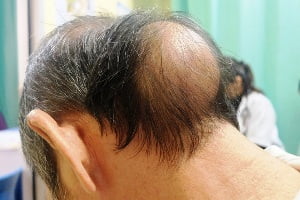
Short-term side effects of radiation therapy may include fatigue, headaches, memory loss, scalp irritation, hair loss, upset stomach and neurologic symptoms and mild skin reactions. Side effects generally depend on the type and dose of the radiation you receive.
Chemotherapy for brain cancer
Chemotherapy is the use of anti cancer drugs to kill the tumour cells. These drugs usually interfere and restrict the cancer cells’ ability to grow and divide. Different objectives of the chemotherapy for brain cancer are:
- to destroy the remaining tumour cells after surgery,
- to slow a tumour’s growth, or
- to reduce the symptoms due to brain tumours
Chemotherapy is generally administered in the form of a capsule or pill which is swallowed orally or via an injection into a vein. This oral or intravenous administration of anti cancer drugs is known as systemic chemotherapy which gets into the bloodstream to reach tumour cells throughout the body. Chemotherapy as a treatment of brain cancer is typically given after surgery and possibly with or after radiation therapy, particularly if the tumour has come back after the initial treatment.
Chemotherapy has a fixed regimen, or schedule, which consists of a specific number of cycles given over a set period of time. During chemotherapy, you may receive 1 drug at a time or a combination of different drugs given at the same time.
Targeted drug therapy
Targeted drug treatments primarily focus on specific genes or proteins of the tumour and the tissue environment present within cancer cells that contributes to a tumour’s growth and survival. Targeted therapy blocks the growth and spread of tumour cells while limiting the damage to healthy cells.
Targeted therapy drugs, in addition to standard chemotherapy are available for certain types of brain tumours, and many more are being studied in clinical trials.
For effective use of targeted therapy, a target must be identified specific to that particular tumour because all tumours do not have the same targets while some may have more than one target. Many research studies are being carried out to find about specific molecular targets of the brain tumours and new treatments directed at them.
Anti-angiogenesis therapy is a type of targeted therapy for the treatment of brain tumours. It stops the process of making new blood vessels restricting the delivery of nutrients to the tumour which makes tumour cells to starve and ultimately die.
Radio surgery
It is not a form of surgery as the name suggests but it uses multiple beams of radiations to cause a highly focused form of radiation treatment to kill the tumour cells in a very small area. Individual beams of radiation aren’t particularly powerful but the point where all the beams meet typically at the location of brain tumour receives a very large dose of radiation enough to kill the tumour cells. It is usually done in one session and you can usually go home the same day. There are many techniques for performing this procedure such as Gamma Knife or linear accelerator radiosurgery.
Recovery and after effects of the brain cancer treatment
After you get the treatment for brain cancer, you may experience some lasting effects and associated problems such as seizures, difficulty in walking and speech problems which need to be addressed properly.
Brain cancer can also affect the areas of the brain that control motor skills, speech, vision and thinking. For such affected areas, rehabilitation may be necessary as part of recovery after the treatment. Your doctor may recommend one of the following based on your needs.
- Physical therapy to regain muscle strength and lost motor skills.
- Occupational therapy to help you to perform your daily normal activities.
- Speech therapy to counter speech difficulties.
- In case of school-going children, tutoring may be needed to help them to cope with changes in memory and thinking.
Brain cancer survival rate: What is the survival rate of brain cancer?
Survival rates give a general idea of the outlook (prognosis) for people with a certain type of tumour. Survival rates can’t tell how long you will live or what will happen to you after the diagnosis of the tumour or cancer. These numbers help in better understanding of how likely it is that your treatment will be successful.
5 – Year relative survival rate for some common brain tumours can be represented by the following figures.
(Source: American Cancer Society)
| Type of Tumour | 5-Year Relative Survival Rate | ||
|---|---|---|---|
| Age | |||
| 20-44 | 45-54 | 55-64 | |
| Low-grade (diffuse) astrocytoma | 68% | 44% | 22% |
| Anaplastic astrocytoma | 54% | 32% | 14% |
| Glioblastoma | 19% | 8% | 5% |
| Oligodendroglioma | 88% | 81% | 68% |
| Anaplastic oligodendroglioma | 71% | 61% | 46% |
| Ependymoma/anaplastic ependymoma | 92% | 89% | 86% |
| Meningioma | 87% | 77% | 71% |
You should know that these numbers are projected estimates and do not predict what will happen in individual cases.
Is it possible to prevent brain cancer?
It is impossible to prevent brain tumours by any known means, but there are certain measures which can reduce your risk of developing brain tumours which include:
- Avoiding exposure to pesticides and insecticides
- Avoiding exposure to carcinogenic or cancer causing chemicals
- Quit smoking
- Protect yourself from unnecessary exposure to radiation


