Fetal VSD Ultrasound
- Updated on: Jul 2, 2024
- 3 min Read
- Published on Feb 27, 2020

What is Ventricular Septum Defect (VSD)?
Ventricular Septum Defect (VSD) is a type of congenital heart disease in which the ventricular septum (wall between the two ventricles) does not form correctly, leaving a hole or defect there. In normal development process, the interventricular septum closes before the fetus is born, so that oxygenated blood is kept mixing with the de-oxygenated blood. When the interventricular septum does not close, it may cause higher pressure in the heart because of increased blood flow or reduced oxygen to the body. Learn more about Ventricular Septum Defect.
How Is Fetal VSD Diagnosed?
Detection of fetal VSD is done by cardiac ultrasound (echocardiogram) performed between 18 and 22 weeks of pregnancy. The methods typically adopted by doctors and cardiologists are:
Fetal Echocardiography
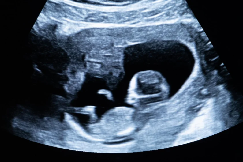
Sound waves (ultrasound) are used in this test to produce a moving image of the heart. The test uses sound waves that echo off the fetal heart structure. This test helps doctors to see abnormalities in the baby’s blood flow and heartbeat.
Fetal electrocardiography is similar to routine pregnancy tests. There are two methods to perform this ultrasound.
Abdominal Echocardiography
If the ultrasound is performed through the abdomen, it’s called an abdominal electrocardiography. While performing this test, a patient has to lie down and expose the belly region of the body. A doctor then applies a lubricating jelly to the skin. This jelly prevents friction so that the doctor can move the ultrasound transducer easily over the skin. The transducer sends and receives sound waves over the skin to capture images.
The jelly sends high frequency sound waves through the body. The waves echo when they hit a dense object inside the body like the fetal heart. Transducer is moved on all over the stomach to obtain images of different parts of the fetal heart. After the procedure, jelly is removed from the abdomen and the patient can return to normal activities.
Transvaginal Echocardiography
If the ultrasound is performed through the vagina, it’s called a transvaginal electrocardiography. In this test, a patient is asked to undress from the waist down and lie on the exam table. Doctor then inserts a probe into the vagina and the probe uses sound waves to create an image of the fetal heart.
Transvaginal electrocardiography is mostly done in the earlier stages of pregnancy and it may provide clear images of the heart.
Techniques Used in Ultrasound Echocardiography
2D-Echocardiography
This technique is used to see the clear structure and motion of heart structures. A 2D echo view appears to be cone-shaped on the monitor allowing motion of the heart structures to be observed. This enables the doctors to evaluate various structures of the heart.
Doppler Echocardiography
This technique is used to measure and analyze blood flow through the heart. The amount of blood pumped out with each beat is a clear indication of the heart functioning.
Color Doppler
It is an enhanced form of Doppler echocardiography. Different colors are used to indicate the direction of blood flow. This clarifies interpretation of the Doppler images.
What Happens After Fetal Electrocardiography?
If the results in an ultrasound are normal, the patient is discharged and asked to revisit if late prenatal tests require it. If a problem is found in the quality of images because of the fetal growth, the patient may need to follow up echo on a later date. If there is a defect in the heart, the doctor may ask for detailed diagnosis and counseling.
Importance of Fetal Echocardiography in the Diagnosis of Ventricular Septal Defect (VSD)
Since heart defects can’t be repaired before delivery, it would be good if they are diagnosed at an early stage so a delivery can be planned. Early diagnosis can help in scheduling the birth of the baby with a neonatal intensive care and to prepare early treatment for the newborn baby such as through a surgical procedure for VSD.


