Mastering the Art of Reading CT Scans for Cancer Diagnosis: Key Tips and Techniques
- Updated on: Feb 29, 2024
- 4 min Read
- Published on Jun 2, 2023

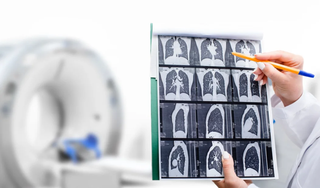
CT scans, or computed tomography scans, are a type of medical imaging that uses X-rays and computer technology to produce detailed images of the body’s internal structures. CT scans are an important tool for cancer diagnosis because they can detect abnormalities such as tumors, masses, and lesions that may not be visible on other types of imaging.
However, interpreting CT scans for cancer diagnosis requires a certain level of skill and expertise. A radiologist or oncologist must be able to accurately identify abnormal growths or tumors, analyze their size, shape, and location, and determine whether they are benign or cancerous. Inaccurate CT scan interpretation can lead to misdiagnosis, delayed treatment, and poor patient outcomes. Therefore, mastering the art of reading CT scans is essential for accurate cancer diagnosis and treatment.
Understanding CT Scans
What is a CT scan? A CT scan uses X-rays and computer technology to create detailed images of the body’s internal structures. During a CT scan, the patient lies on a table that moves through a doughnut-shaped machine that takes X-ray images from multiple angles. The images are then combined by a computer to create cross-sectional images of the body.
How CT scans work by using X-rays to produce detailed images of the body’s internal structures. X-rays are a type of radiation that can pass through the body, but different tissues absorb different amounts of X-rays. The CT scanner detects the X-rays that pass through the body and uses this information to create an image.
Different types of CT scans there are different types of CT scans, including:
- Contrast-enhanced CT scans: These scans use a contrast agent, which is a type of dye that is injected into the patient’s bloodstream. The contrast agent makes certain tissues and structures more visible on the CT scan.
- Virtual colonoscopy: This is a type of CT scan that is used to screen for colon cancer.
Advantages and limitations of CT scans have several advantages, including:
- They are non-invasive and painless
- They produce detailed images of the body’s internal structures
- They can detect abnormalities that may not be visible in other types of imaging
However, CT scans also have limitations, including:
- They expose the patient to radiation
- They may not be able to distinguish between benign and cancerous growths
- They may produce false positives or false negatives
Reading CT Scans for Cancer Diagnosis
Key components of a CT scan report typically include the following components:
- Patient information: This includes the patient’s name, age, and medical history.
- Technique: This describes the type of CT scan that was performed, including any contrast agents that were used.
- Findings: This describes any abnormalities that were detected on the CT scan, including the size, shape, and location of any tumors or masses.
- Impression: This is the radiologist’s or oncologist’s overall interpretation of the CT scan, including any recommendations for further testing or treatment.
Understanding the different types of images in a CT scan CT scans produce different types of images, including:
- Axial images: These are cross-sectional images of the body that are taken in a horizontal plane.
- Sagittal images: These are images that are taken in a vertical plane and show the body from front to back.
- Coronal images: These are images that are taken in a vertical plane and show the body from side to side.
Identifying abnormal growths or tumors Radiologists and oncologists must be able to identify abnormal growths or tumors on a CT scan. This may involve comparing the CT scan to previous scans, looking for areas of abnormal density, and assessing the size, shape, and location of any masses.
Analyzing the size, shape, and location of tumors Analyzing the size, shape, and location of tumors is an important part of CT scan interpretation for cancer diagnosis. The radiologist or oncologist will measure the size of any tumors and assess their shape and location in relation to other structures in the body. This information can help determine the stage of the cancer and guide treatment decisions.
Identifying metastatic tumors are tumors that have spread from their original location to other parts of the body. Identifying metastatic tumors on a CT scan can be challenging, as they may be small or located in hard-to-see areas. Radiologists and oncologists must carefully examine the CT scan for any signs of metastatic tumors and may need to use other imaging techniques, such as MRI or PET scans, to confirm their presence.
Tips for Accurate CT Scan Interpretation
Importance of Communication Between Radiologists And Oncologists
Communication between radiologists and oncologists is essential for accurate CT scan interpretation. Radiologists must provide clear and concise reports that are easy for oncologists to understand, and oncologists must provide radiologists with relevant patient information and guidance on the interpretation of the CT scan.
Use of Contrast Agents
Contrast agents can be useful for enhancing the visibility of certain tissues and structures on a CT scan. However, their use must be carefully considered, as they can also produce false positives or cause allergic reactions in some patients.
Consideration of the Patient’s Medical History
The patient’s medical history can provide important context for CT scan interpretation. For example, a patient with a history of lung cancer may be more likely to develop lung metastases, which would influence the interpretation of a CT scan of the chest.
Importance of Reviewing Previous CT Scans
Reviewing previous CT scans can provide important information for CT scan interpretation, such as whether a tumor has grown or changed over time. Radiologists and oncologists should always review previous scans when available.
Staying Up-To-Date With the Latest Research and Technology
Advances in CT scan technology and research are constantly improving our ability to detect and diagnose cancer. Radiologists and oncologists should stay up-to-date with the latest developments in CT scan interpretation to ensure the best possible patient outcomes.
Challenges in CT Scan Interpretation
Variability in radiologist interpretation Interpreting CT scans is a subjective process, and different radiologists may interpret the same scan differently. This can lead to variability in diagnosis and treatment decisions, which can impact patient outcomes.
Overcoming technical difficulties CT scans can be affected by technical difficulties such as motion artifacts, which can produce inaccurate images. Radiologists and technicians must be trained to recognize and overcome these difficulties to ensure accurate CT scan interpretation.
Dealing with false positives and false negatives CT scans can produce false positives, which are abnormal findings that turn out to be benign, or false negatives, which are missed abnormalities that are later discovered to be cancerous. Radiologists and oncologists must be aware of the limitations of CT scans and use them in conjunction with other imaging techniques and clinical information to minimize the risk of false positives and false negatives.
Mastering the art of reading CT scans is essential for accurate cancer diagnosis and treatment. Radiologists and oncologists must be able to accurately identify abnormal growths or tumors, analyze their size, shape, and location, and determine whether they are benign or cancerous. By following the key tips and techniques outlined in this article, and staying up-to-date with the latest research and technology, radiologists and oncologists can ensure the best possible patient outcomes.


