Salivary Gland Cancer – An Overview
- Updated on: Jul 12, 2024
- 18 min Read
- Published on Oct 3, 2019

Salivary Gland Cancer – Definition and Overview
Earlier, salivary gland cancer used to be considered as one of the rarest types of cancers in human body. But, recently it is becoming much more common. A salivary gland tumor begins in the salivary glands. These glands are found in neck, mouth or throat regions. When malignant cancer cells develop in the form of tissues on the salivary glands, it marks the beginning of salivary gland cancer.
This cancer is one of the main cancers of head and neck region. It metastasizes to other parts of the body through lymphatic (blood) system relatively easily.
What are Salivary glands?
The salivary glands comprise of tissues. The function of these glands is to produce saliva and keep the mouth moist. Saliva has a few enzymes that help in the digestion (chew, swallow and digest) of food. It also contains antibodies which kill infections in mouth and throat.
Check out our graphics for salivary gland cancer: Salivary gland cancer images and photographs.
Types of salivary glands
There are two types of salivary glands:
- Major salivary glands
- Minor salivary glands
Major salivary glands
There are three main pairs of salivary glands which are supposed to be major salivary glands and are found under or behind the jaw in the mouth. They are:
The parotid glands
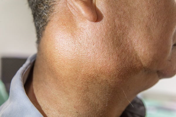
Parotid glands are the largest glands found in-front or just under the ear lobes on both sides of the face. Approximately, 70% of the tumors begin from parotid glands and most of them are benign.
The sublingual glands
Sublingual glands are located under the tongue (below the jaw). These glands are smaller in size and have more chances of malignancy. Approximately, 15% of the tumors arise from these glands.
The submandibular glands
Tumors rarely begin from submandibular glands. Submandibular glands are the smallest of the three major salivary glands. They are placed under the tongue at the base of the mouth (each side of the jawbone).
Minor salivary gland
There are hundreds (about 600) of types of salivary glands in or around the mouth. Some are so tiny that they are only visible through microscope. They are present in lips, inside the cheeks, windpipe, throughout mouth and throat (area of the upper jaw along the inside of the teeth and the soft palate), and parts of the tissue lining near the upper digestive tract (known as the mucosa).
Types of salivary gland tumors
Mainly, salivary gland tumors are classified into two types: Benign and Malignant.
Benign salivary gland tumors
Benign salivary gland tumors are not life-risking cancers. They do not spread to other parts of the body. Most salivary gland tumors develop in the parotid salivary gland and are benign in nature.
Benign salivary gland tumors are often cured when they start budding rapidly. The best way to cure benign tumors is surgery. If these tumors are not treated for a larger duration, they can become highly cancerous. They can often grow back (recur) if not treated completely.
Common types of benign salivary gland tumors are adenomas (monomorphic adenoma), oncocytomas, Warthin tumors, benign mixed tumors (or pleomorphic adenomas) and papillary cystadenoma lymphomatosum (earlier it was known as cylindroma).
Malignant salivary gland tumors
Malignant tumors in a salivary gland are comparatively less common than benign tumors. Salivary gland cancers which are malignant are described on the basis of their cell type (as they appear under the microscope).
Grading of salivary gland cancer
The type of a malignant salivary gland cancer is described by its grade (G). Grading helps to provide an idea about how cells multiply and compares the health of cancer cells to normal cells when viewed under a microscope.
The grading system of salivary gland cancer is described below:
- Grade 1 (also called low grade or well differentiated)
The cells in this grade are slow growing and appear similar to normal salivary gland cells. They have good outcome (prognosis).
- Grade 2 (also called intermediate grade or moderately differentiated)
The cancer cells look abnormal compared to the cells in Grade 1.
- Grade 3 (also called high grade or poorly differentiated
The cancer cells tend to grow more quickly and look very different (abnormal) from the normal cells. The outlook for these cancers is usually not as good as for lower grade cancers.
Types of salivary gland cancers
Mucoepidermoid carcinoma
Mucoepidermoid carcinoma is the most common type of low grade-salivary gland cancer. They more often begin in parotid glands.
Adenoid cystic carcinoma
Adenoid cystic carcinoma is also a low grade salivary gland cancer which slowly grows and becomes malignant. It is very difficult to completely treat adenoid cystic carcinoma as it spreads along the nerves.
Adenocarcinomas
Adenocarcinomas begin in the gland cells. The main types of adenocarcinomas are:
- Acinic cell carcinoma
Acinic cell carcinomas start in the parotid gland and tend to grow slowly. They occur at a young age and are low graded.
- Polymorphous low-grade adenocarcinoma (PLGA)
Polymorphous low-grade adenocarcinoma initiates from minor salivary glands and are mostly curable. They might not always grow slow.
- Other adenocarcinomas are: Adenocarcinoma, Basal cell adenocarcinoma, Clear cell carcinoma, Cystadenocarcinoma, Sebaceous adenocarcinoma, Sebaceous lymphadenocarcinoma, Mucinous adenocarcinoma, not otherwise specified (NOS) etc.
Malignant mixed tumors
There are 3 types of malignant mixed tumors:
- Carcinoma ex pleomorphic adenoma
- Carcinosarcoma
- Metastasizing mixed tumor
Some rare salivary gland cancers
- Squamous cell carcinoma
SCC develops easily in older men and has poor outlook.
- Epithelial-myoepithelial carcinoma
EMC is a low grade cancer and has a tendency to reoccur.
- Anaplastic small cell carcinoma
Anaplastic cancer has a tendency to grow quickly and is found in minor salivary glands.
- Non-Hodgkin lymphoma
Non-Hodgkin lymphoma is a cancer that spreads in immune cells in or near the salivary glands leading to Non-Hodgkin Lymphoma.
- Secondary salivary gland cancer
Secondary salivary gland cancers begin in any other organ and spreads to the salivary gland from there.
Causes of Salivary gland cancer, Salivary gland cancer risk factors
The exact cause of salivary gland cancer is still unknown. But there are a few risk factors as listed below which are considered to increase the chances of someone developing salivary gland cancer. However, not everyone with these risk factors develop the cancer. Similarly, not having any of these risk factors does not mean that someone will never develop it.
Age and Gender
It was observed that in about 75% of cases, an increase in age raises the risk of salivary gland cancer. Older people suffer more with salivary gland cancer. It was found that it is more common in males than in females.
Radioactive substance exposure
Radiotherapy of the head and neck to treat any other cancer increases the chances of getting salivary gland cancer. Children are at a greater risk of salivary gland cancer during radiotherapy.
Occupational or workplace environment
Exposure to chemicals, sawdust, pesticides, and different industrial solvents leads to increased risk of salivary gland cancer (in nose and sinuses). Salivary gland cancer can be caused by an exposure to certain radioactive substances in workplaces such as in rubber manufacturing, mining of asbestos or in chemical industries and plumbing.
Family history
Although it is rare, but people with a family history of salivary gland cancer have higher chances of getting the disease.
Other possible risk factors
These factors include lifestyle and dietary habits of a person. It may be possible that a diet which is low in vegetables and high in animal fat may increase the risk of salivary gland cancer. A study found an increased risk of parotid gland tumors among heavy cell-phone users. The use of tobacco and alcohol raises the risk of several types of cancers of the head and neck area.
How much do we know about the causes of salivary gland cancer?
The various risk factors for getting salivary gland cancer do not indicate the exact cause of the disease. It is usually not an inherited cancer, and therefore the changes that occur in a person due to the cancer may be quite different in different persons.
These random changes in a person might be due to exposure to radioactive elements, or chemicals, or lifestyle of a person. Salivary gland cancer might occur when mutations or changes develop in the DNA of some cells in a salivary gland. This in turn allows the cells to grow, divide and die. These cells together form a mass, resulting in a cancerous cluster which spreads to mouth and neck and then to different organs. There is not enough evidence to prove that these are the exact causes of salivary gland cancer.
Prevention of salivary gland cancer: Can salivary gland cancer be prevented?
As the exact cause for salivary gland cancer is not known, prevention of the cancer is also not possible completely. However, once can minimize the risk by minimizing the risk factors for salivary gland cancer such by avoiding radioactive exposure, tobacco use, consumption of excess alcohol and by living a healthy lifestyle.
Taking precautions might help in lowering the risk of salivary gland cancer but it may not always prevent it.
Screening and early detection of salivary gland cancer: Can salivary gland cancer be detected at an early stage?
Although, the symptoms of salivary gland cancer are not very common or specific but depending on its location, the salivary gland cancer can be easily detected.
If a lump appears within one of the salivary glands (usually on the sides of the face or in the mouth), diagnosing the tumors in the salivary glands may become easy as part of general routine medical and dental check-ups. Common signs and symptoms of the cancer should not be ignored though they may overlap with other health conditions and diseases. Early stage detection can help in delivering the right treatment.
Signs and symptoms of salivary gland cancer
Most benign and malignant tumors are often painless masses in the beginning. There are a few signs and symptoms, however, which indicate the start of tumors in the salivary glands. Sometimes, the symptoms can appear due to something else.
The signs and symptoms of salivary gland cancer are:
- A lump or swelling which is usually painless (in mouth, cheek, jaw, or neck)
- Pain in the mouth, cheek, jaw, ear, or neck which does not go away
- Numbness in parts of the face
- Weakness of the muscles on one side of the face
- Trouble opening mouth widely
- Fluid draining from ear
- Trouble swallowing
- Malignant tumors can cause pain, numbness, loss of motor function of neurons in nerves, etc by invading nerves.
Diagnosing salivary gland cancer
The diagnosis of salivary gland cancer depends on the symptoms which become visible in the patient with course of time. Most salivary gland tumors are benign, therefore most of the times the symptoms do not show up in the beginning.
Many times, routine checkups help in the diagnosis of a salivary gland tumor. If a salivary gland cancer is detected, the distant organs are also checked for metastasis.
There are a few tests and procedures for diagnosing salivary gland cancer after the symptoms are visible. Some of them are:
Physical examination and medical history
Studying the medical history is supposed to be the first and foremost step in the diagnosis of salivary gland cancer.
The signs and symptoms of salivary gland cancer are carefully viewed and tests are done for further evaluation. If the results of these tests are abnormal, doctors refer you for complete treatment as defined based on the stage of your salivary gland cancer.
During a physical examination, the lumps, bumps or any unusual thing such as swollen jaw, throat or neck are detected. These are the most important signs of any abnormality in salivary glands. Facial paralysis or dentals problems are also an indication of the salivary gland cancer.
Imaging tests for salivary gland cancer
Imaging tests such as MRI, CT scan, X-ray help in the detection cancer as well as determining the exact size and location of the tumor in the salivary gland by creating images of the tumor. Images are also collected from distant sites to check if the tumor has metastasized to other locations.
Some of the commonly performed imaging tests for diagnosing salivary gland cancer are:
MRI (Magnetic Resonance Imaging) for salivary gland cancer
Magnetic resonance imaging (MRI) uses powerful magnetic fields (instead of X-rays) to produce images of soft tissues in the body. Nuclear Magnetic Resonance Imaging (NMRI) is the procedure which uses magnet and radio-waves to produce computerized detailed images of the body.
A special dye (contrast medium) called gadolinium is injected in the body before the test. The dye helps in visualization of the cancer and differentiating between the tumors and the unaffected part. Enlarged lymph nodes can also be easily detected through an MRI test.
CT scan (computerized tomography scan) for salivary gland cancer
CT scan is also known as computerized axial tomography scan. 3D images of the organs (soft tissues) inside the body are created using X-rays by capturing different angles of the body. These images at different angles provide a cross sectional view that depicts any abnormalities or tumors. The shape, size and position of a tumor are clearly defined in these images.
- CT scan can also help to locate enlarged lymph nodes that might contain salivary gland cancer. It also shows images of other organs that have suspicious spots which might be due to metastasized salivary gland tumors.
- The contrast dye is either injected in the vein (intravenously) or given as a liquid (oral contrast) to be swallowed (orally).
Positron emission tomography (PET) scan for salivary gland cancer
- PET is one of the finest tests to diagnose malignant tumor cells in the body. Mostly, a combination of PET and CT (PET-CT) scan is used to detect salivary gland cancer.
- Instead of looking for areas with an abnormality (depending upon the size and shape), the PET-CT scan images the areas with high cellular activity (which might be cancerous).
- A small quantity of radioactive glucose (sugar) is injected intravenously in the patient’s body. This radioactive glucose is taken up by cells that use the most energy (as the cancer cells tend to utilize energy actively by absorbing more amount of radioactive sugar than normal cells).
- A rotating scanner detects the radioactive sugar in the body and produce images of portions inside the body. Malignant salivary gland cancer cells appear to be brighter than normal cells.
Ultrasound for salivary gland cancer
Sound waves are used to detect salivary gland cancer. Results of an ultrasound are supposed to be superficial. Ultrasonic images help us understand the size and pattern of the salivary gland cancer.
Endoscopy for salivary gland cancer
A thin, flexible tube-like instrument with a light and a lens for viewing called endoscope is inserted though the mouth, down the esophagus into the stomach and the bowel.
The examination of tumors with endoscopy may involve such as laryngoscopy (for examining larynx), pharyngoscopy (for examining pharynx), or a nasopharyngoscopy (for examining nasopharynx).
Biopsy for salivary gland cancer
Biopsy is a commonly used method for diagnosing cancers. In a biopsy procedure, a small amount of tissue is removed from the tumor mass (affected area) and is viewed under a microscope. This method helps detecting the actual condition of the cells. There are certain methods to perform biopsy as follows:
Fine needle aspiration (FNA) biopsy
- A small quantity of cells in the form of liquid is removed from the suspected lump. A thin hollow needle is inserted into the body and a little fluid is extracted which is tested in a microbiology laboratory to detect the presence of cancerous cells.
- For certainty, FNA is used for the detection of salivary gland cancer.
- In many cases, FNA helps prevent unnecessary surgery.
- FNA also defines whether a lump is due to an infection or if it is a benign cancer tumor.
Incisional biopsy
- A very small part of the lump is taken and sent to a laboratory for the detection of salivary gland cancer.
- It is done in cases when the FNA samples are not enough.
- It is also called Open biopsy
Panendoscopy
A series of connected tunes are used to look through the following organs to diagnose salivary gland cancer:
- pharynx (throat)
- larynx (voice box)
- oesophagus (food pipe)
- trachea (windpipe)
- bronchi (breathing tubes)
A panendoscope is inserted through the nose to the throat carefully with anaesthesia. It is not a common diagnosis method for salivary gland cancer.
Stages of salivary gland cancer
The stage of a cancer shows how big it is and whether it has spread to nearby and/or distant locations. The stages of a salivary gland cancer are defined on the basis of the results of a physical exam, imaging tests and biopsy. A cancer stage describes the size of the tumor and locates the distant sites where the salivary gland cancer has extended.
T: It describes the size of tumor and tells whether or not it has metastasized.
N: N explains the degree of tumor spread to the lymph nodes.
M: M defines whether the tumor in the salivary gland has spread to distant sites such as lung or liver.
Stage 1 salivary gland cancer: Tis, N0, M0
The tumor (Tis) is confined to cells lining of salivary duct. Tis stands for tumor or carcinoma in situ.
It has neither spread to the lymph nodes (N0) nor it has metastasized to different organs of the body (M0).
Stage 2 salivary gland cancer: T1, N0, M0
The tumor of salivary gland is of 2 cm or smaller in size. It doesn’t grow into nearby tissues (T1) and doesn’t affect the lymph nodes (N0). It causes no effect to other organs of the body (M0).
Stage 3 salivary gland cancer: T3, N0, M0 or T0, T1, T2, T3, N1, M0
Stage 3 constitute two conditions –
First condition: T3, N0, M0
or
Second condition: T0, T1, T2, T3, N1, M0
In the first condition, the tumor in the salivary gland is more than 4 cm in size and continues to grow into nearby soft tissues (T3). At this stage, there is no development of tumor in the lymph nodes (N0) and other organs of the body (M0).
In the second condition, the cancer grows into the nearby soft tissues and can be of any size (T0-T3) and spreads to 1 lymph node (N1) on the same side of the head or neck as the primary tumor. The cancer has not grown outside of the lymph nodes. The salivary gland cancer has not spread to the distant sites (M0).
Stage 4A salivary gland cancer
T4a, N0 or N1, M0
The salivary gland cancer can be of any size and grows into nearby structures such as the jaw bone, skin, ear canal, or facial nerve. This is known as moderately advanced disease (T4a). The tumor has either not spread or has spread to 1 lymph node (N0 or N1). It has not grown outside of the lymph node (not more than 3 cm in size). It does not spread to distant sites (M0).
OR
T0/T1/ T2/ T3 or T4a, N2a/N2b/N2c, M0
The salivary gland cancer grows into any size and grows into nearby soft tissues or structures such as the jaw bone, skin, ear canal, and/or facial nerve (T0-T4a).
The tumor grows into one side of the lymph node. It has not grown outside the lymph nodes.
- The tumor grows into one side of the lymph node. The size of the tumor is not more than 6cm (N2a, N2b).
- The tumor might not enlarge more than 6cm but can spread to either on both sides of the neck or at the back of the neck (N2c)
- The salivary gland cancer has not spread to distant sites (M0).
Stage 4b salivary gland cancer
Any T, N3, M0
The tumor in the salivary gland can be of any size and might have grown into nearby soft tissues or structure (Any T).
The tumor spreads to the lymph nodes, either it is larger than 3cm and outside the lymph node on opposite side or both sides of the lymph nodes (N3b) or larger than 6cm but not outside the lymph node (N3a) or it has spread opposite side of the primary cancer which is 3 cm or smaller and has grown outside of the lymph node (N3c).
The tumor has not spread to distant organs (M0).
OR
T4b, Any N, M0
The tumor can be of any size and grows into nearby structures such as the base of the skull or other bones nearby, or it surrounds the carotid artery. The disease is at “very advanced” stage (T3b).
The tumor may or may not spread to nearby lymph nodes (Any N) and to distant organs (M).
Stage 4c salivary gland cancer
Any T, Any N, M1
The tumor is of any size and grows into nearby soft tissues or structures (Any T). It may or may not have spread to nearby lymph nodes (Any N). The tumor metastasizes at this stage to distant organs such as the lungs.
The following additional categories of stages are not listed above:
TX: The main tumor cannot be assessed due to lack of information.
T0: Primary tumor is not visible.
NX: Accession to regional lymph node is not possible due to lack of information.
Treatment of salivary gland cancer
The treatment of salivary gland cancer depends on several factors:
- The type of salivary gland cancer
- The location of tumor
- The stage and grade of the cancer
- Impact of treatment on speech, chewing and swallowing
- Lifestyle and age of the patient
Some of the commonly used treatment options for salivary gland cancer are:
Surgery for salivary gland cancer
Surgery is the first line of treatment which ensures complete removal of the tumor from the salivary glands and the surrounding areas to which it has spread. Some healthy tissues, called margins, around the tumor are also removed. Surgery can be done in four different ways based on how the tumor affects your body:
- Removing the tumor from salivary gland
If the tumor is small in size and visible then it becomes easy for the surgeon to detect it and remove it along with some surrounding healthy tissues.
- Remove the complete salivary gland
Entire salivary gland and the connected organs are also removed if the tumor has spread to a larger area.
- Removal of lymph nodes near the salivary gland
If there is any possibility that the tumor of the salivary gland has affected the lymph nodes, then these lymph nodes are removed through surgery (of neck).
- Reconstructive surgery
After removing the salivary glands, the reconstruction of that area is recommended to repair and maintain the normal functioning of body such as chewing, speaking, swallowing or breathing. Grafting is the way to reconstruct the removed portions of the neck and mouth.
Types of surgery for salivary gland cancer
Parotid gland surgery
This is the largest and major salivary gland which has two lobes (superficial lobe – outside part of the gland and deep lobe – deeper tissues) present on either side of the mouth and in front of both ears. Its function is to produce saliva which has digestive properties and has antibacterial properties. It also lubricates the mouth.
- Superficial parotidectomy: Superficial parotidectomy mainly involves surgery of the superficial lobe in case of low grade cancers
- Total parotidectomy: The complete parotid gland is removed during total parotidectomy.
Submandibular or sublingual gland surgery
This type of surgery involves complete removal of submandibular gland and two sublingual glands with some tissues and lymph nodes which might be affected with salivary gland cancer. These glands control the movement of tongue, facial expressions, sensation and taste.
Minor salivary gland surgery
The size and location describe the treatment option for salivary gland cancer. In a minor salivary gland surgery, small salivary glands located throughout the lining of the mouth and throat are removed if affected.
Reconstructive surgery
Reconstructive (plastic) surgery is used to rebuilt tissue and nerves of mouth and jaw that were removed during surgery to eliminate the salivary gland cancer. The main aim of reconstructive surgery is to help a person eat, drink and speak normally and maintain the appearance.
Lymph node removal (neck dissection)
The removal of cancer-affected lymph nodes through surgery is called lymph node dissection or lymphadenectomy. It is also known as neck dissection surgery.
A neck dissection surgery is done when:
- Lymph nodes are enlarged (in the neck)
- The lymph nodes may contain salivary gland cancer
- The salivary gland cancer is high-grade and involves high risk of spreading
The after-effects of neck dissection surgery can result in numbness of the ear, weakness when raising the arm above the head, and weakness of the lower lip.
Endoscopic surgery
Endoscopic surgery is a less destructive surgery to healthy tissues for removing salivary gland tumor especially in the paranasal area (around the nose) or in the larynx. However, it is rarely done.
Side effects of salivary gland cancer surgery
Neck being a sensitive part of the body can be affected by the surgery. The nerves near the salivary glands may get damaged which may lead to uncontrolled facial expression, voice defect, numbness in ear, weakness in raising arm above the head, and weakness of the lower lip.
Radiation therapy for salivary gland cancer
Radiation therapy uses strong energy beams (X-rays or protons) which destroy the cancer cells or lower down their growth. This therapy can be used alone or can be clubbed with surgery if there is any chance of spread of salivary gland cancer in any other part of the body.
Types of radiation therapy used are:
- External beam radiation therapy: The beams focus from outside the body to treat salivary gland cancer. These beams with the required dosage are angled in such a way that they shall not affect any other body part except the target region.
- Three-dimensional conformal radiation therapy (3D-CRT): 3D-CRT uses imaging scan results to mark location of the tumor. The beams of required shape are aimed at the tumor from different directions. These radiation beams are of low intensity and therefore do not damage any tissues, but collectively they provide a high dose of radiation at the tumor region.
- Intensity modulated radiation therapy (IMRT): IMRT is an advanced form of 3D therapy which also involves shaping of the beams and aiming them at the affected region.
- Internal radiation therapy: Internal radiation is also known as Brachytherapy. It uses implants such as tiny rods or pellets which contain radioactive pellets that are delivered in or nearby the tumor.
- Fast neutron therapy: Fast neutron therapy is successful in treating salivary gland tumors especially the ones that are unresectable. Neutron radiation therapies mechanize a beam of high-energy neutrons which is significantly more effective than photons.
Possible side effects of radio-therapy
- Major side effect of radiation therapy is reduced saliva, which can lead to a dry mouth
- Slight redness or darkening occurs on the other side of the body where the radiotherapy beams leave the body
- A patient feels weak and lacking energy, but this might gradually improve.
- Difficulty in swallowing, hearing, dental problems, hair loss, etc.
Chemotherapy for Salivary Gland Cancer
The use of anti-drugs to kill the cancer cells and reduce their growth is the basic principle of chemotherapy. These drugs are given intravenously or orally.
If radiotherapy or surgery does not cure the salivary gland cancer, chemo drugs are used to shrink the tumor. These drugs do not cure the cancer completely but restricts the spread of cancer cells which affect other nearby organs.
If chemo drugs are given along with the radiotherapy, it is known as chemo-radiation therapy. Some of the chemo drugs used in the treatment of salivary gland cancers include:
- Cisplatin
- Doxorubicin (Adriamycin®)
- Cyclophosphamide (Cytoxan®)
- Docetaxel (Taxotere®)
- Methotrexate
- Paclitaxel (Taxol®)
- 5-fluorouracil (5-FU)
- Carboplatin
- Vinorelbine (Navelbine®)
Side effects of chemotherapy
The side effects of chemotherapy depend upon:
- The type of drug
- The amount of drug
- Body’s reaction to the drug
Common chemotherapy side effects include:
- feeling sick or fatigue
- hair loss
- bleeding and bruising easily
- appetite loss
- feeling tired / weakness
- lower resistance towards infections
- weight loss
- diarrhoea or constipation
- damage to nerves (called neuropathy) leading to burning or tingling sensations, sensitivity to cold or heat, pain, etc
Treatment Options by Stage of Salivary Gland Cancer
Treatment of Stage 1 salivary gland cancer
Surgery with or without radiation therapy is recommended for low grade cancer. Radiation therapy is generally advised after surgery during an intermediate- or high-grade salivary gland cancer, if it cannot be completely eliminated through the body.
A few cells (edges or signs) are left as symptoms if it is not completely treated. Participating in clinical trials is also another option for treating salivary gland cancer.
Treatment of Stage 2 salivary gland cancer
Salivary gland cancer might extend to lymph nodes but is still restricted in the nearby area. Therefore, surgery and radiation therapy are again the preferred treatment options.
Fast neutron or photon-beam radiation therapy is done for high grade salivary gland cancer at stage 2.
Treatment of Stage 3 salivary gland cancer
Salivary gland cancers have started to grow outside the salivary gland which might have also reached the lymph nodes in the neck.
The treatment at this stage includes the following:
- Surgery with lymphadenectomy followed by radiation therapy is the most basic treatment plan.
- Fast neutron radiation therapy is also given to reduce the growth of cancer cells.
- Participating in clinical trials of radiation therapy and/or radio-sensitizers and chemotherapy might be an option at this stage
Treatment of Stage 4 salivary gland cancer
Stage 4 has no cure in most cases. All the treatment options can be considered to increase the lifespan for the patient. The tumor is burnt to shrink so that the growth of cancer cells gets slow. Clinical trials are conducted especially on stage 4 patients to seek any benefit for them.
Prognosis, outlook and survival rates for salivary gland cancer by stage
Salivary gland cancer is one of the rarest types of cancers. The 5-year survival rate which is studied on the percentage of people (number of survivors out of 100 patients) surviving the cancer for salivary gland cancer is 72%. The average age of patients taken into account is about 60 years.
People diagnosed with cancer of the major salivary glands between 1998 and 1999 provided these statistical data depending upon the stage of salivary gland cancer:
Stages and their 5-year Relative Survival Rate:
Stage1- 91% survival rate for 5 years
Stage 2- 75% survival rate for 5 years
Stage 3- 65% survival rate for 5 years
Stage 4- 39% survival rate for 5 years
Recurrent salivary gland cancer
There is a great probability that once a person has had cancer, he or she may get it again even after the successful treatment. It is also known as “second cancer”. Sometimes, the cancer might not hit the same organ but any other organ.
People who survive the salivary gland cancers have an increased risk of:
- Another salivary gland cancer (more malignant and different from that of the first time)
- Thyroid cancer
- Cancer of the oral cavity (mouth)
- Lung cancer
What happens if the treatment fails at any stage of salivary gland cancer?
Salivary gland cancer affects a very sensitive part of the body. One of the sensory organs of the body i.e. tongue is highly affected due to this type of cancer.
The treatment is stage-dependent but if the chances of recovery are very less, it can be very stressful for patients. The lifestyle of a patient can increase or decrease the effect of the treatment going on.
Salivary gland cancer often affects other organs and spreads relatively easily. Therefore, it is important to ensure right treatment is delivered before the tumor cells metastasize to other organs and lymph nodes.


