The Retina of Human Eye: Definition, Function, Anatomy
- Updated on: Aug 3, 2024
- 4 min Read
- Published on Apr 22, 2021


Retina Definition
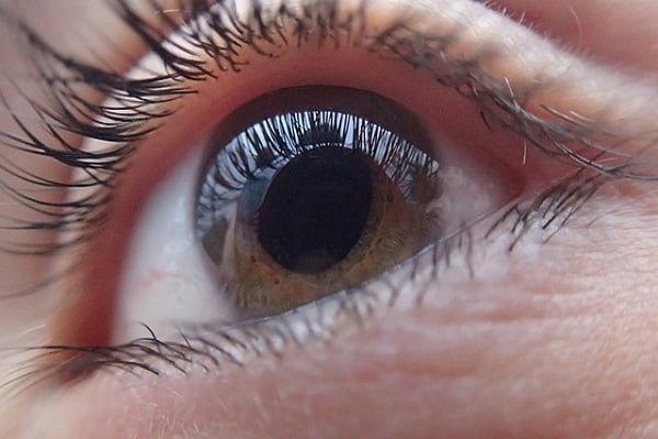
The retina of human eye is the sensory membrane that lines the inner surface of the back of the eyeball. It contains several layers, including a layer that contains photoreceptors.
Our retina is about the size of a postage stamp. It consists of a central area called the macula and a larger peripheral area of the retina. Retina is made of a layer of nervous tissue that covers the inside of about two-thirds of the eyeball at back. It is in fact an extension of your brain, formed from neural tissue and connected to the brain by the optic nerve.
Anatomy and Structure of Retina

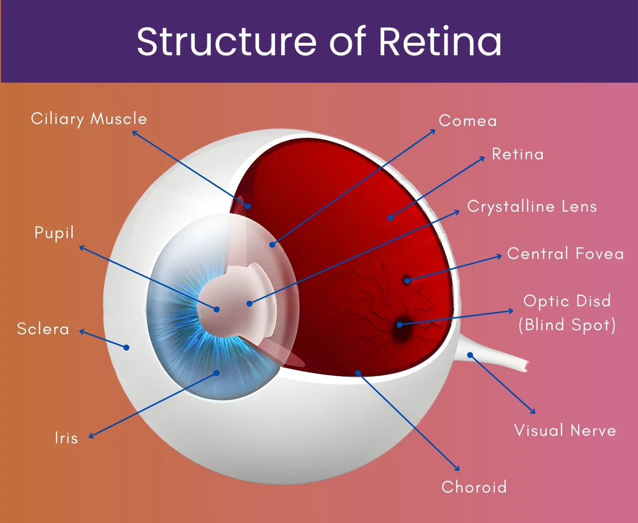
Human eye retina is a complex tissue that contains many layers. Out of these several layers, only one layer contains light-sensitive photoreceptor cells.
Light must pass through the overlying layers to reach the photoreceptor cells. Photoreceptors are of two types:
- Rods or rod cells
- Cones or cone cells
The various parts of retina are discussed herein.
Rods or rod cells
Rod cells are photoreceptor cells in the retina of the eye that can work in less intense light. Rods are usually found at the outer edges of your retina and are responsible for peripheral vision. Rods function mainly in dim light and provide black-and-white vision.
There are about 90 million rod cells in the retina of a human eye. These cells are more sensitive than cone cells and are almost entirely responsible for night vision. But, rods have very limited role in color vision. This is the reason why colors are not very much clear in the night or in darkness. Rods are a little longer than cones but the basic structure is almost same.
Cones or cone cells
Cone cells, or cones, are one of three types of photoreceptor cells in the retina of eyes. Cone cells are primarily responsible for your color vision. These cells function in relatively bright light, as opposed to rod cells, which work better in dim light such as in the night.
There are primarily three types of cone cells according to our current understanding of the retina:
- Red cone cells
- Green cone cells
- Blue cone cells
It is believed that there are about 6 to 7 million cones that can be divided into the above three types as follows::
- red cones – 64%
- green cones – 32%
- blue cones – 2%
Cone cells provide color sensitivity to our eyes. The green and red cones are concentrated in the fovea centralis. The blue cones are mostly found outside the fovea. Blue cones have the highest color sensitivity.
The rod and cone cells are differentiated structurally by their characteristic shapes and their functionality (their sensitivity to different kinds of light).
Rods are well distributed over the entire retina, but cones are usually concentrated at:
- the fovea centralis (at the rear of the retina)
- surrounding macula lutea
See also: Ishihara’s Test For Color Deficiency (Color Blindness)
See also: Retinal Detachment: Causes, Symptoms, Diagnosis, Treatment
Macula
The macula is a small central area of the retina that contains a high concentration of cones. It allows clear central vision to see fine details for activities such as viewing small objects from a distance, reading a book, threading a needle, etc.
In the center of the macular is the fovea. The fovea is considered to be the most important part of the eye. Generally, vision is not lost until the fovea is affected by diseases such as diabetic retinopathy or macular degeneration etc. Damage to macula is the most common cause of blindness.
Light is focused by various structures in the front of the eye such as cornea & lens at the macula. It takes the picture of an object that is sent to the brain, where vision is formed. The macula provides us the ability to read and see in fine details, and the rest of the retina provides peripheral vision. In humans, macula has a high degree of sophisticated and complex structure.
Function of the Retina
Photoreceptor cells of the retina accept light focused by the cornea and lens and convert it into chemical and nervous signals which are sent to the visual center of our brain through the optic nerve.
These signals are converted into images in the visual cortex of the brain, located at the back of the brain.
When light enters your eye, it first enters through the cornea and the lens and is refracted to focus an image onto the retina. Light-sensitive chemicals in the rods and cones react to it and cause nerve impulses to generate signals. These impulses are combined together through a complex system of interconnections in retinal cell layers to generate a coherent pattern. This is then transmitted to the brain through the optic nerve, where they are further interpreted. Thus, an image is seen and visual perceptions are formed.
In short, our retina basically processes a picture from the focused light and sends it to the brain, which then decides what the picture is.
Since a retina plays a vital role in image formation and recognition, a damage or disruption to it can cause complete blindness.
Common diseases and conditions of the retina are listed below.
More: Anti–Vascular Endothelial Growth Factor Therapy (Anti VEGF Therapy) For Eye Disorders And Cancers
More: Diabetic Retinopathy: Causes, Symptoms, Diagnosis, Treatment, Complications
Diseases and conditions of the retina
- Macular degeneration
- Diabetic retinopathy
- Retinal detachment
- Retinoblastoma
- Retinal Tear
- Epiretinal membrane
- Macular hole
- Retinitis pigmentosa
Common symptoms indicative of a problem with your retina
- Lost vision – partially or completely
- Sudden onset of blurred or distorted vision – straight lines look away
- Defects in the side vision
- Eye floaters along with flashes
- Seeing floating specks
- Shadows or blind spots in the field of vision


1 Comment
I would be happy to participate in the study. Many years ago, I had a detached retina but my eye doctor had a way to reattach the retina. I called what he did as a kind of “soldering” job. Recently, I had the cornea in my right eye removed and replaced with a new cornea which gives me GREAT vision.
If you want me, in the study I will be available.