Understanding Your ECG for Pericarditis
- Updated on: Jun 25, 2024
- 3 min Read
- Published on Mar 3, 2020

What do You Mean by Pericarditis?
Pericardium is a thin, double membranous sac in which the heart is enclosed. The inflammation of pericardium produces chest pain, dyspnoea and tachycardia. This condition is called pericarditis.
There may be friction between the layers of pericardium due to the leakage of blood or fluid, which as a result may become inflamed. That inflamed tissue rub against each other if you have pericarditis.
Pericardial effusion and cardiac temponade are the causes of pericarditis. Acute pericarditis is a type of inflammatory pericarditis in which chest pain develops suddenly. Check out the causes of pericardial effusion.
How is Pericarditis Diagnosed?
Pericarditis is diagnosed through electrocardiograph (ECG), echocardiography, magnetic resonance imaging (MRI), and computed topography (CT).
What is Pericarditis Electrocardiography (ECG)?
Electrical activity of the heart is recorded by electrocardiography (ECG). A graph of voltage versus time is recorded as waves with different patterns corresponding to each electrical activity of the heart in an ECG.
ECG can also be done to check irregular heartbeats, shortness of breath, high blood pressure, and heart valve problems.
An ECG constitutes of three waves:
P wave: It represents atrial depolarization
QRS wave: It reflects ventricular depolarization
T wave: It represents ventricular repolarisation
How is ECG Performed for Diagnosing Pericarditis and other Heart Problems?
ECG is a painless and non-invasive test with quick results. In a conventional electrocardiogram 12-lead ECG, 10 electrodes (sensors) are attached on a patient’s limbs and surface of the chest to determine the electrical activity of heart. In this way, the magnitude of a heart’s electric potential is measured from twelve different angles.
The patient has to lie flat with the head and chest raised a little. The patient is asked to relax for a few minutes during an ECG procedure so that the electrodes can be placed on the body properly to give reliable results. These sensors are left on a patient’s body for only a few minutes.
Stages of Pericarditis ECG
Pericarditis ECG changes occur in these progressive stages, all of which can be seen in almost 50% pericarditis cases:
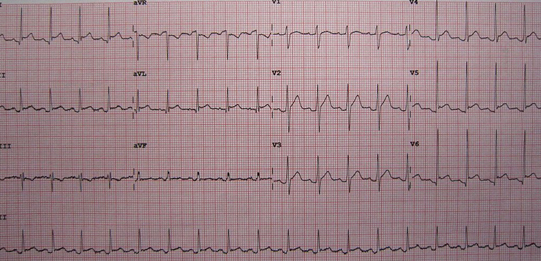
Stage I: This stage is also known as acute pericarditis and is recognized by diffuse ST elevation and PR elevation in most of the leads and periodically notching at the QRS complex end.
Stage II: It is featured by resolvation of ST segment elevation, PR depression and T waves may get flattened. This is also termed as pseudo normalization.
Stage III: Only T wave inversion is found in this stage.
Stage IV: This is characterized by a return of T wave to the upright position and normalization of ECG.
How Can Pericarditis ECG be Distinguished With Early Repolarisation ECG?
There are three things on which pericarditis can be distinguished with early polarization:
- In early repolarisation, ratio of T wave amplitude to the ST elevation must be greater than 4, which means early repolarization T wave is usually 4 times the amplitude of ST segment elevation.
- ST elevation in pericarditis ECG cannot be resolved by performing exercises.
- Pericarditis patients are found to have symptoms typical of pericarditis such as chest pain etc, but early repolarisation is not associated with any symptoms.

(Image Credit:Wikimedia Commons)
The figure above shows a typical ECG of a pericarditis patient.
FAQs
Why is There ST Elevation in Pericarditis ECG?
Electrocardiography changes in the pericarditis leads to diffused ST elevation in most of the electrodes of the 12 lead electrocardiograms. ST elevation in pericarditis ECG is associated with the inflammatory changes in the pericardium underlying the inflamed pericardium of heart. The electrical signal from this area of ST elevation must return to some unaffected region of the heart and this should be linked with the ST depression region.
What Triggers Pericarditis?
Pericarditis can be caused by bacterial, parasitic, and fungal infection. Autoimmune diseases like scleroderma, rheumatoid arthritis can also trigger pericarditis. Kidney failure and myocardial infarction can also leads to pericarditis.
Does Pericarditis Cause Damage to the Heart?
Pericardium is a thin, sac like double membranous structure which encloses the heart. In pericarditis, this pericardium becomes inflamed. Although this inflammation is mild and transient but it may lead to severe illness and prolonged inflammation can cause cardiac damage.
What Causes PR Depression in ECG?
ST elevation can be found in pericarditis but there should be no reciprocal ST depression (except V1 and AVR). PR depression may also be a cause of myocardial infarction (due to atria infarction).


