What is ventricular septal defect?
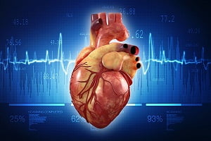
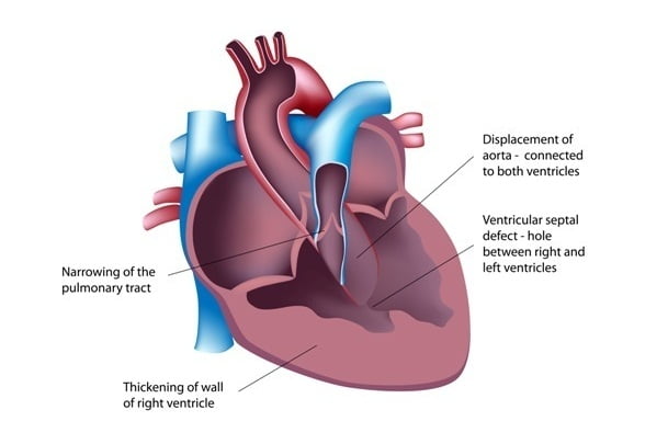
Ventricular septal defect is a birth defect (congenital) of the heart in which there is a hole in the wall (septum) that divides the lower two chambers (ventricles) of the heart. It results due to incomplete formation of the wall during fetal development stage. This separating wall is also known as ventricular septum.
This “hole” – sepatal defect, often allows mixing of oxygen-rich blood from the left ventricle to the right ventricle and back to the lungs instead of out to the body as it happens in normal conditions, causing the heart to work harder along with pulmonary congestion.
Ventricular septal defects occur in many locations and sizes. This defect often occurs on its own or with other congenital heart diseases.
A small ventricular septal defect poses no life threatening problems, as many of small VSDs close on their own with time. Medium and larger VSDs may need surgical repairs early in life to prevent increased risk of other complications including but not limited to:
- heart failure
- high blood pressure in the lungs (Pulmonary Hypertension
- Irregular heart rhythms (such as Arrhythmia)
- Stroke
What are the types of ventricular septal defect?
Although there are several classifications for VSDs based on the size of defect, location of the defect, number of defects and presence or absence of ventricular septal aneurysm – a harmless thin flap of the tissue on the septum.
According to most used classification system, there are 4 basic types of VSD:
Membranous VSD
This is an opening located in the upper part of the ventricular septum, near the aortic and tricuspid valves. This type of VSD doesn’t usually close spontaneously, therefore a surgery is often needed.
Muscular VSD
This is an opening in the muscular portion of the lower section of the ventricular septum. Most of these muscular VSDs close spontaneously and do not need surgery.
Atrioventricular canal type VSD
This type of VSD is associated with atrioventricular canal defect. It is located just next to the tricuspid and mitral valves. It needs surgical repair.
Conal septal VSD
It is of the rarest kind of VSD located in the ventricular septum just below the pulmonary valve.
What are the signs and symptoms of ventricular septal defect?
Ventricular septal defect is usually symptomless at birth. It usually starts manifesting symptoms after a few weeks of birth.
The size of the opening in the ventricular septum and other associated heart defects determine the severity of symptoms, and the age at which they first appear. Larger openings allow greater amount of blood to pass through and overload the right ventricles and lungs.
Following are the most common symptoms of VSD observed in a baby. However, each child may experience symptoms differently.
Symptoms may include:
- Fatigue
- Sweating
- Rapid breathing
- Heavy breathing
- Congested breathing
- Disinterest in feeding or tiring while feeding
- Poor weight gain/ failure to thrive
- Frequent respiratory infections
Doctor may first suspect a heart defect during a regular check-up if he or she hears a distinct whooshing sound called heart murmur while listening to your baby’s heart with a stethoscope.
Sometimes VSD is not detected until a person reaches adulthood.
What are the causes of ventricular septal defect?
Most ventricular septal defects occur by chance with no defined etiology or with no clear reason of their occurrence. It arises from the problems in early heart development.
During fetal development, a ventricular septal defect occurs due to incomplete formation of a muscular wall (septum) which divides the heart into left and right, between the lower chambers of the heart.
VSD are also thought to be due to a combination of genetics and other factors such as environment, the things mother comes in contact to or due to medicines which mother uses.
It is possible to acquire VSD later in life, usually after a heart attack or myocardial infarction or as a result of certain heart procedures.
What are the risk factors for ventricular septal defect?
VSD often occurs at the same time with other birth defects. Many of the factors which increase the risk of other defects also pose a risk of VSD.
Specific risk factors include:
- Being of Asian heritage
- Having a family history of congenital heart disease
- Having other genetic disorders such as Down syndrome
- Alcohol consumption
- Using antiseizure medicines such as Dilantin during pregnancy
How is ventricular septal defect diagnosed?
Your child’s doctor will go for a physical exam after sign and symptoms of any heart defect are manifested.
Ventricular septal defects are always associated with a characteristic murmur sound, which is simply a sound caused by the passage of blood through the opening from left to right side of the heart. The location where this sound is best heard along with loudness and quality of the murmur give the doctor an initial idea about the heart defect. He or she might suggest some of these tests to confirm the diagnosis:
Echocardiogram/Echocardiography:
In this test, sound waves (called ultrasound) are used to produce a moving picture of the heart. Doctors use this test to diagnose a ventricular septal defect and to determine its location, size and severity.
The test may also be used to detect any other heart problems and how heart is reacting to the problems.
Echocardiography can be used on a fetus (fetal echocardiography) as well. This test will help a cardiologist decide whether and when the treatment is needed.
Electrocardiogram (ECG):
It is a simple, painless diagnostic test which records the heart’s electrical activity with the help of electrodes attached to the skin. It also records and reflects the strength and timing of the electrical signals as they pass through different parts of the heart.
Using this test, a cardiologist determines heart defects or rhythm-related problems.
Chest X Ray:
It is used to look if there is an enlarged heart and if there is extra fluid in the lungs.
Cardiac catheterization:
A cardiac catheterization is an invasive procedure where a thin, flexible tube (catheter) is inserted into a blood vessel at the groin or arm region and guided through the blood vessels into the heart.
Special dye is injected through the catheter into a blood vessel or a chamber of the heart. The dye allows the doctor to clearly visualise the flow of the blood through the heart and the blood vessels on an x-ray image.
The doctor also can use cardiac catheterization to estimate the pressure inside the heart chambers and the blood vessels. This can help the doctor determine whether blood is mixing between the two sides of the heart and to what extent.
Pulse Oximetry:
It shows how much oxygen is there in the blood by using a small sensor attached to the finger tip or toe like an adhesive band.
How is a ventricular septal defect treated/cured?
If the VSD is small and without any symptoms, a wait and watch approach is employed to see if the defect reverses itself and if the hole closes properly and if there is no further risk of heart failure.
Some types of VSD may need to be corrected if it has not closed on its own to prevent lung problems that will develop from a continuous exposure to the extra blood flow.
When treatment for a VSD is required, options are such as extra nutrition and surgery to close the VSD.
Babies who have large VSDs and tire easily during feeding may require extra nutrition consisting of high calorie to help them grow.
Here are some options of treatment that are available.
Medications for ventricular septal defect
Some children will need medication to help strengthening their heart muscles, control high blood pressure and decrease the extra fluid in the circulation and the lungs.
Surgical and other Procedures for the treatment of ventricular septal defect
In more severe cases, where ventricular septal defect is larger and has failed to respond medication or symptoms for a heart failure is manifested, doctors generally recommend for surgery. In such cases, a VSD is closed either with stitches or patches in an open-heart surgery.
Another approach known as Catheter procedure is employed to close the opening in septum where a incision in chest is not required instead a thin tube (catheter) is inserted into the blood vessel in the groin and guided to the heart. The doctor uses a special-sized mesh to close the hole.
Most of the surgeries are performed using these two procedures either alone or in combination.
What are the complications of ventricular septal defect?
A small VSD may never create any problem but medium and large VSDs can have severe effects ranging from mild to life threatening complications.
Possible complications of VSD may include:
- Aortic insufficiency (leaking of the valve that separates the left ventricle from the aorta)
- Delayed growth and development
- Heart failure
- Infective endocarditis – Bacterial infection of the heart
- Pulmonary hypertension
- Other heart problems which may include heart rhythm and valve problems
How can ventricular septal defect be prevented?
Except for the VSD which is caused by a heart attack, this condition is always present at birth. There is nothing you can do to prevent having a baby born with ventricular septal defect. But every precaution should be practised to have a healthy pregnancy.
Some of them include:
- Get prenatal counselling even before you get pregnant with your doctor
- Eat a balanced diet
- Exercise regularly
- Avoid risks such as alcohol, tobacco and illegal drugs
- Avoid having infections
- Keep diabetes under control – consult your doctor and be sure it’s under control before you get pregnant
