Browsing: Ventricular Septal Defect
Comprehensive Information, Resources, and Support on Ventricular Septal Defect
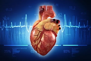
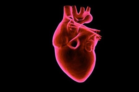
Human Heart: Anatomy, Function, Chambers, Location, Facts
The heart is a muscular organ that works as your body’s circulatory pump. It takes in deoxygenated blood through the veins and delivers it to the lungs for oxygenation and then pumps this oxygenated blood into the arteries. In coronary heart disease, the heart does not function properly. Learn more about your heart.
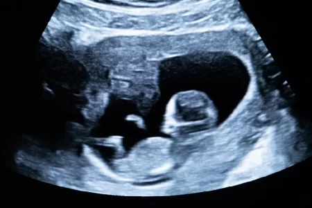
Fetal VSD Ultrasound
Detection of fetal VSD is done by cardiac ultrasound performed between 18 and 22 weeks of pregnancy. Sound waves (ultrasound) are used in this test to produce a moving image of the heart. The test helps doctors to see abnormalities in a baby’s blood flow and heartbeat.
Many a times untreated VSD is diagnosed in adults and it may require surgery if it can cause a risk to the patient. In adults, VSD can be small, medium, or large in size with or without other complications such as pulmonary stenosis, pulmonary hypertension, or aortic regurgitation.
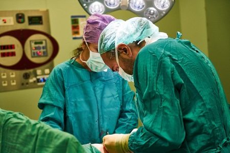
A Ventricular Septal Defect (VSD) surgery is a type of heart surgery which is performed to correct a hole between the left and right ventricles. Severe complications after the surgery are rare and the VSD surgery success rate is very high. You should ask your child’s doctor about risks after a VSD surgery.
